. 2010 Jul;7(7):535-40.
doi: 10.1038/nmeth.1468. Epub 2010 Jun 6.

Two-photon calcium imaging from head-fixed Drosophila during optomotor walking behavior

Affiliations

Two-photon calcium imaging from head-fixed Drosophila during optomotor walking behavior

Johannes D Seelig et al. Nat Methods. 2010 Jul.

Erratum in

  • Nat Methods. 2011 Feb;8(2):184

Abstract

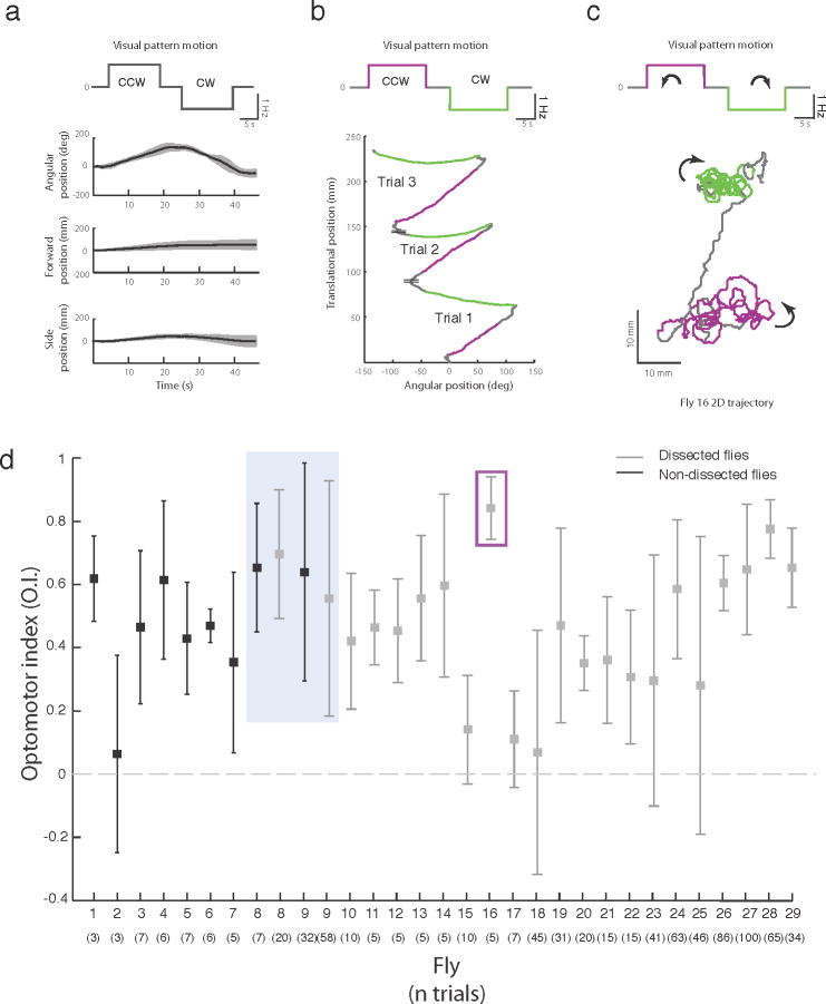
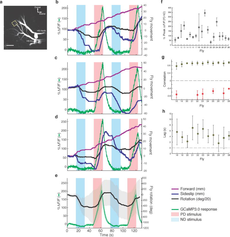
Drosophila melanogaster is a model organism rich in genetic tools to manipulate and identify neural circuits involved in specific behaviors. Here we present a technique for two-photon calcium imaging in the central brain of head-fixed Drosophila walking on an air-supported ball. The ball's motion is tracked at high resolution and can be treated as a proxy for the fly's own movements. We used the genetically encoded calcium sensor, GCaMP3.0, to record from important elements of the motion-processing pathway, the horizontal-system lobula plate tangential cells (LPTCs) in the fly optic lobe. We presented motion stimuli to the tethered fly and found that calcium transients in horizontal-system neurons correlated with robust optomotor behavior during walking. Our technique allows both behavior and physiology in identified neurons to be monitored in a genetic model organism with an extensive repertoire of walking behaviors.


Conflict of interest statement

Competing interests statement

The authors declare no competing financial interests.

Figures

Figure 1
Figure 1. Setup for two-photon imaging from the brain of head-fixed flies walking on a ball
(a) Fly holder for tethered walking fly recording separates exposed brain from intact legs and eyes allowing visual stimulation and walking on the ball. (b) Schematic showing arrangement of holder, ball, ball trackers, calibration camera, microscope (including a schematic for 2-photon excitation and detection systems), objective and visual arena.
Figure 2
Figure 2. High-precision ball tracking system allows online measurement of fly’s virtual trajectory
(a) Both cameras capture X and Y velocity in their respective fields of view. Together, they provide 4 kHz tracking of the ball’s rotation about all three axes. The brown arrow shows the fly’s direction on the ball. (b) Rotational velocity of the ball can be converted to the tethered fly’s translational and rotational velocities on a virtual 2D surface. (c), (d) In an exemplary trajectory, the instantaneous velocity (5 ms bins) of a walking fly is shown along with the accumulated displacement and rotation of the fly. (eh) Fly displacement can be plotted as a virtual trajectory on a flat surface. Sample trajectories shown are examples of spontaneous walking without motion stimuli. Color tracks passage of time. Velocities for trajectory (e) are shown in (c).
Figure 3
Figure 3. Optomotor behavior in tethered flies
(a) Rotational and translational velocity of one fly in response to clockwise (CCW) and counterclockwise (CW) motion stimuli. The fly’s rotational velocity, plotted as mean ± s.d., n = 5 trials, tracks the direction of the motion stimulus. (b) An exemplary trajectory showing the translational and rotational movement of the fly color-coded to emphasize the stimulus switches from CCW to CW. (c) Virtual trajectory of one fly in response to the motion stimulus reveals optomotor response as CCW and then CW loops (see also SI movies M1 and M2). (d) Summary plot showing mean ± s.d. (n for each provided in Supplementary Fig. 4c) of optomotor indices across trials for both non-dissected and dissected flies. Trials in gray box show pre- and post-dissected optomotor performance for the same flies. Trials highlighted in magenta are those shown in (b).
Figure 4
Figure 4. Optical imaging of dendrites of a motion-sensitive neuron during tethered walking
(a) Typical Z-projected image of HS-neurons expressing GCaMP3.0 (R27B03-Gal4). Only HS-North and HS-Equatorial are clearly visible. The rectangle outlines a typical distal dendritic region of interest selected for imaging. Scale bar: 20 μm. (b-d) Examples of single trials showing HS-neuron calcium transients and Fly 24’s simultaneously recorded behavioral response as the motion stimulus alternates from the HS-neuron’s ND to PD. Note that rotation is shown in degs/20 for ease of plotting with forward and sideslip movement. (e) Mean ± s.d. of seven trials of Fly 24’s accumulated rotation (in degs) and HS-neuron calcium transients during motion stimulation. (f) Mean± s.d. (n for each in Supplementary Fig. 4c) of difference between peaks of PD and ND calcium transients across trials for all flies (variability mainly reflects differences between imaging in different regions of interest). (g) Peak correlations between calcium responses of HS neurons and rotational response to PD stimulation for Protocol 2 flies and trials with positive O.I. (see Online Methods for details). Means for occasional trials with negative correlations are shown in red (numbers of trials available in Supplementary Information). (h) Lags across trials and flies measured from onset of calcium response to onset of behavioral rotational response for trials with positive correlation. All flies with the exception of Fly 20 show lags significantly greater than zero (t-test, p-values < 0.05; all p-values available in Supplementary Information).

Similar articles

Cited by

References

    1. Vosshall LB. Into the mind of a fly. Nature. 2007;450:193–197. - PubMed
    1. Olsen SR, Wilson RI. Cracking neural circuits in a tiny brain: new approaches for understanding the neural circuitry of Drosophila. Trends Neurosci. 2008;31:512–520. - PMC - PubMed
    1. Borst A. Drosophila’s view on insect vision. Curr Biol. 2009;19:R36–47. - PubMed
    1. Simpson JH. Mapping and manipulating neural circuits in the fly brain. Adv Genet. 2009;65:79–143. - PubMed
    1. Gouwens NW, Wilson RI. Signal propagation in Drosophila central neurons. J Neurosci. 2009;29:6239–6249. - PMC - PubMed

Publication types

LinkOut - more resources