

Engineered disulfide bonds restore chaperone-like function of DJ-1 mutants linked to familial Parkinson's disease
- PMID: 20527929
- PMCID: PMC2925438
- DOI: 10.1021/bi902164h
Engineered disulfide bonds restore chaperone-like function of DJ-1 mutants linked to familial Parkinson's disease
Abstract
Loss-of-function mutations such as L166P, A104T, and M26I in the DJ-1 gene (PARK7) have been linked to autosomal-recessive early onset Parkinson's disease (PD). Cellular and structural studies of the familial mutants suggest that these mutations may destabilize the dimeric structure. To look for common dynamical signatures among the DJ-1 mutants, short MD simulations of up to 1000 ps were conducted to identify the weakest region of the protein (residues 38-70). In an attempt to stabilize the protein, we mutated residue Val 51 to cysteine (V51C) to make a symmetry-related disulfide bridge with the preexisting Cys 53 on the opposite subunit. We found that the introduction of this disulfide linkage stabilized the mutants A104T and M26I against thermal denaturation, improved their ability to scavenge reactive oxygen species (ROS), and restored a chaperone-like function of blocking alpha-synuclein aggregation. The L166P mutant was far too unstable to be rescued by introduction of the V51C mutation. The results presented here point to the possible development of pharmacological chaperones, which may eventually lead to PD therapeutics.
Figures
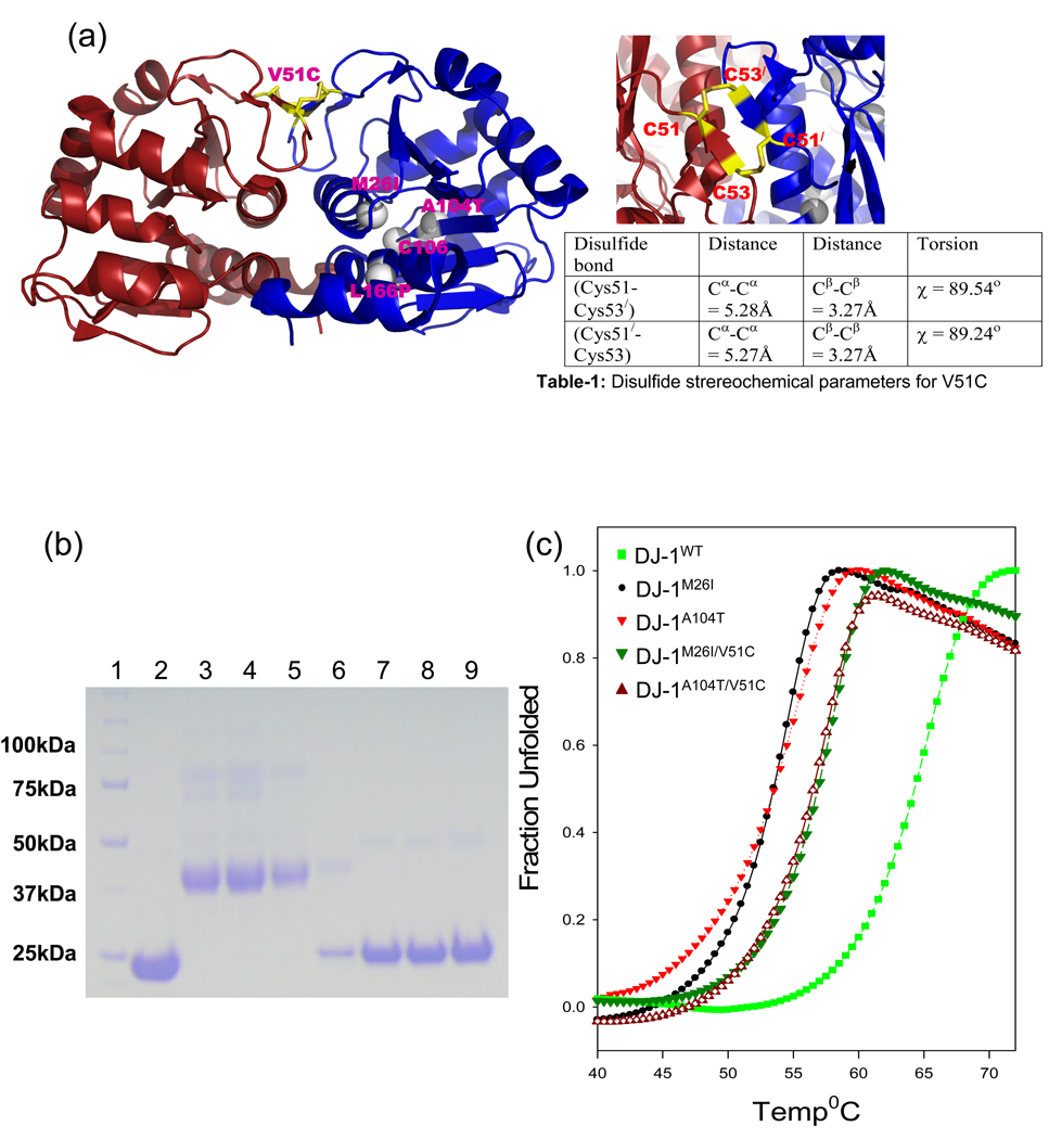


 , p<0.0001 Student’s t-test). Similarly, DJ-1M26I/V51C showed higher glutathione peroxidase activity as compared to DJ-1M26I (n=5,
  , p=0.0004 Student’s t-test). (b) Aggregation of α-synuclein in presence of various DJ-1WT and various mutants. Data from five independent aggregation experiments for α-synuclein incubated various forms of DJ-1 was used for generation of the plot and error bars. α-synuclein aggregates to completion within 48hrs under our assay conditions. The mutants DJ-1A104T and DJ-1M26I have no effect on α-synuclein aggregation kinetics. DJ-1WT delays the aggregation of α-synuclein with the first Thio-T positive aggregates appearing around 80hrs. The V51C disulfide bonded mutants DJ-1A104T/V51C and DJ-1A104T/V51C delays the aggregation of α-synuclein with the first Thio-T positive aggregates appearing after 60hrs.
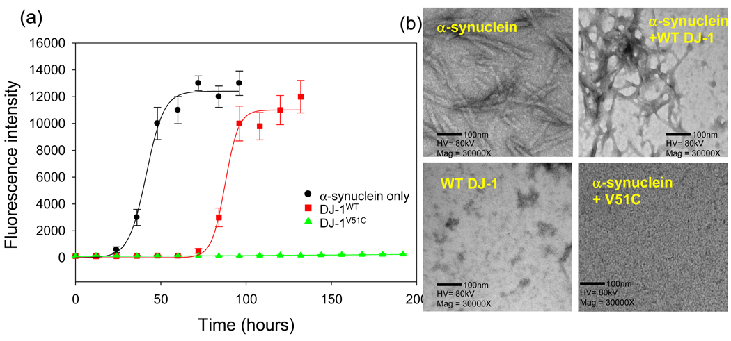
References
-
- Dawson TM, Dawson VL. Molecular pathways of neurodegeneration in Parkinson's disease. Science. 2003;302:819–822. - PubMed
-
- Bonifati V, Rizzu P, van Baren MJ, Schaap O, Breedveld GJ, Krieger E, Dekker MC, Squitieri F, Ibanez P, Joosse M, van Dongen JW, Vanacore N, van Swieten JC, Brice A, Meco G, van Duijn CM, Oostra BA, Heutink P. Mutations in the DJ-1 gene associated with autosomal recessive early-onset parkinsonism. Science. 2003;299:256–259. - PubMed
-
- Macedo MG, Anar B, Bronner IF, Cannella M, Squitieri F, Bonifati V, Hoogeveen A, Heutink P, Rizzu P. The DJ-1 L166P mutant protein associated with early onset Parkinson's disease is unstable and forms higher-order protein complexes. Hum Mol Genet. 2003;12:2807–2816. Epub 2003 Sep 2802. - PubMed
-
- Miller DW, Ahmad R, Hague S, Baptista MJ, Canet-Aviles R, McLendon C, Carter DM, Zhu PP, Stadler J, Chandran J, Klinefelter GR, Blackstone C, Cookson MR. L166P mutant DJ-1, causative for recessive Parkinson's disease, is degraded through the ubiquitin-proteasome system. J Biol Chem. 2003;278:36588–36595. Epub 32003 Jul 36588. - PubMed
-
- Abou-Sleiman PM, Healy DG, Quinn N, Lees AJ, Wood NW. The role of pathogenic DJ-1 mutations in Parkinson's disease. Ann. Neurol. 2003;54:283–286. - PubMed
Publication types
MeSH terms
Substances
Grants and funding
LinkOut - more resources
Full Text Sources
Other Literature Sources
Medical
Miscellaneous
