

Resting cerebral blood flow alterations in chronic traumatic brain injury: an arterial spin labeling perfusion FMRI study
- PMID: 20528163
- PMCID: PMC2967826
- DOI: 10.1089/neu.2009.1215
Resting cerebral blood flow alterations in chronic traumatic brain injury: an arterial spin labeling perfusion FMRI study
Abstract
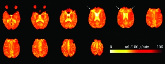
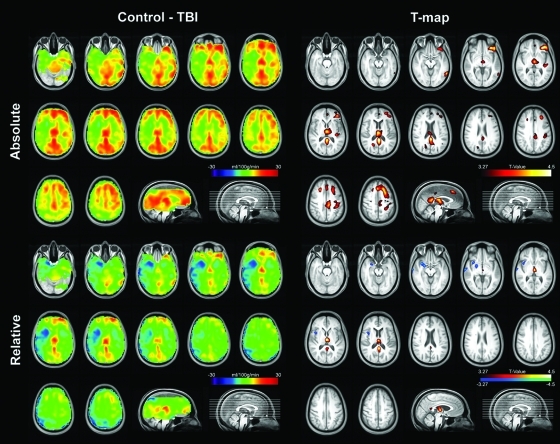
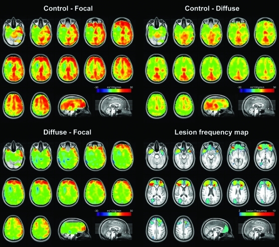
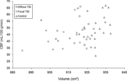
Non-invasive measurement of resting state cerebral blood flow (CBF) may reflect alterations of brain structure and function after traumatic brain injury (TBI). However, previous imaging studies of resting state brain in chronic TBI have been limited by several factors, including measurement in relative rather than absolute units, use of crude spatial registration methods, exclusion of subjects with substantial focal lesions, and exposure to ionizing radiation, which limits repeated assessments. This study aimed to overcome those obstacles by measuring absolute CBF with an arterial spin labeling perfusion fMRI technique, and using an image preprocessing protocol that is optimized for brains with mixed diffuse and focal injuries characteristic of moderate and severe TBI. Resting state CBF was quantified in 27 individuals with moderate to severe TBI in the chronic stage, and 22 demographically matched healthy controls. In addition to global CBF reductions in the TBI subjects, more prominent regional hypoperfusion was found in the posterior cingulate cortices, the thalami, and multiple locations in the frontal cortices. Diffuse injury, as assessed by tensor-based morphometry, was mainly associated with reduced CBF in the posterior cingulate cortices and the thalami, where the greatest volume losses were detected. Hypoperfusion in superior and middle frontal cortices, in contrast, was associated with focal lesions. These results suggest that structural lesions, both focal and diffuse, are the main contributors to the absolute CBF alterations seen in chronic TBI, and that CBF may serve as a tool to assess functioning neuronal volume. We also speculate that resting reductions in posterior cingulate perfusion may reflect alterations in the default-mode network, and may contribute to the attentional deficits common in TBI.
Figures
Similar articles
-
The relationship of resting cerebral blood flow and brain activation during a social cognition task in adolescents with chronic moderate to severe traumatic brain injury: a preliminary investigation.Int J Dev Neurosci. 2012 May;30(3):255-66. doi: 10.1016/j.ijdevneu.2011.10.008. Epub 2011 Nov 22. Int J Dev Neurosci. 2012. PMID: 22120754 Free PMC article.
-
Arterial Spin Labeling Reveals Elevated Cerebral Blood Flow with Distinct Clusters of Hypo- and Hyperperfusion after Traumatic Brain Injury.J Neurotrauma. 2021 Sep 15;38(18):2538-2548. doi: 10.1089/neu.2020.7553. Epub 2021 Jun 10. J Neurotrauma. 2021. PMID: 34115539 Free PMC article.
-
Dynamic association between perfusion and white matter integrity across time since injury in Veterans with history of TBI.Neuroimage Clin. 2016 Dec 23;14:308-315. doi: 10.1016/j.nicl.2016.12.017. eCollection 2017. Neuroimage Clin. 2016. PMID: 28210542 Free PMC article.
-
Advanced neuroimaging in traumatic brain injury: an overview.Neurosurg Focus. 2019 Dec 1;47(6):E17. doi: 10.3171/2019.9.FOCUS19652. Neurosurg Focus. 2019. PMID: 32364704 Review.
-
Perfusion Imaging of Traumatic Brain Injury.Neuroimaging Clin N Am. 2023 May;33(2):315-324. doi: 10.1016/j.nic.2023.01.006. Neuroimaging Clin N Am. 2023. PMID: 36965948 Review.
Cited by
-
Mapping the Connectome Following Traumatic Brain Injury.Curr Neurol Neurosci Rep. 2016 May;16(5):44. doi: 10.1007/s11910-016-0642-9. Curr Neurol Neurosci Rep. 2016. PMID: 27021773 Review.
-
Functional magnetic resonance imaging during emotion recognition in social anxiety disorder: an activation likelihood meta-analysis.Front Hum Neurosci. 2013 Jan 17;6:347. doi: 10.3389/fnhum.2012.00347. eCollection 2012. Front Hum Neurosci. 2013. PMID: 23335892 Free PMC article.
-
Dynamic Functional Network Analysis in Mild Traumatic Brain Injury.Brain Connect. 2019 Jul;9(6):475-487. doi: 10.1089/brain.2018.0629. Brain Connect. 2019. PMID: 30982332 Free PMC article.
-
The Insight ToolKit image registration framework.Front Neuroinform. 2014 Apr 28;8:44. doi: 10.3389/fninf.2014.00044. eCollection 2014. Front Neuroinform. 2014. PMID: 24817849 Free PMC article.
-
Network dysfunction after traumatic brain injury.Nat Rev Neurol. 2014 Mar;10(3):156-66. doi: 10.1038/nrneurol.2014.15. Epub 2014 Feb 11. Nat Rev Neurol. 2014. PMID: 24514870 Review.
References
-
- Ances B.M. Zarahn E. Greenberg J.H. Detre J.A. Coupling of neural activation to blood flow in the somatosensory cortex of rats is time-intensity separable, but not linear. J. Cereb. Blood Flow Metab. 2000;20:921–930. - PubMed
-
- Anderson K.E. Taber K.H. Hurley R.A. Functional imaging. In: Silver J.M., editor; McAllister T.W., editor; Yudofsky S.C., editor. Textbook of Traumatic Brain Injury. American Psychiatric Publishing; Washington DC: 2005. pp. 107–133.
-
- Avants B. Gee J.C. Geodesic estimation for large deformation anatomical shape averaging and interpolation. Neuroimage. 2004;23(Suppl. 1):S139–S150. - PubMed
-
- Azouvi P. Neuroimaging correlates of cognitive and functional outcome after traumatic brain injury. Curr. Opin. Neurol. 2000;13:665–669. - PubMed
Publication types
MeSH terms
Substances
Grants and funding
LinkOut - more resources
Full Text Sources
Medical
