. 2010 Jun;5(4):563-74.
doi: 10.2217/nnm.10.30.

Enhanced binding and killing of target tumor cells by drug-loaded liposomes modified with tumor-specific phage fusion coat protein

Affiliations

Enhanced binding and killing of target tumor cells by drug-loaded liposomes modified with tumor-specific phage fusion coat protein

Tao Wang et al. Nanomedicine (Lond). 2010 Jun.

Abstract

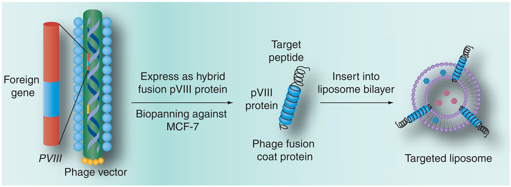
Aim: To explore cancer cell-specific phage fusion pVIII coat protein, identified using phage display, for targeted delivery of drug-loaded liposomes to MCF-7 breast cancer cells.

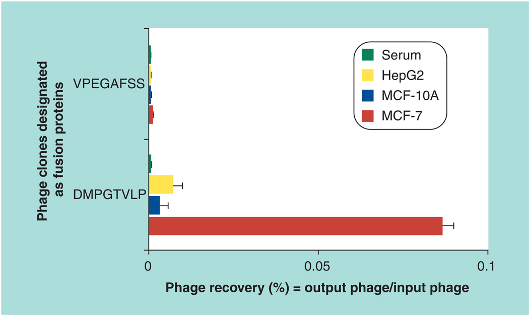
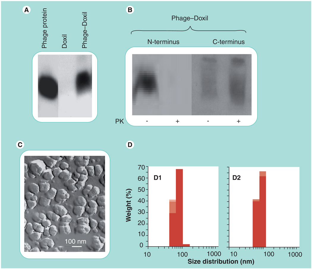
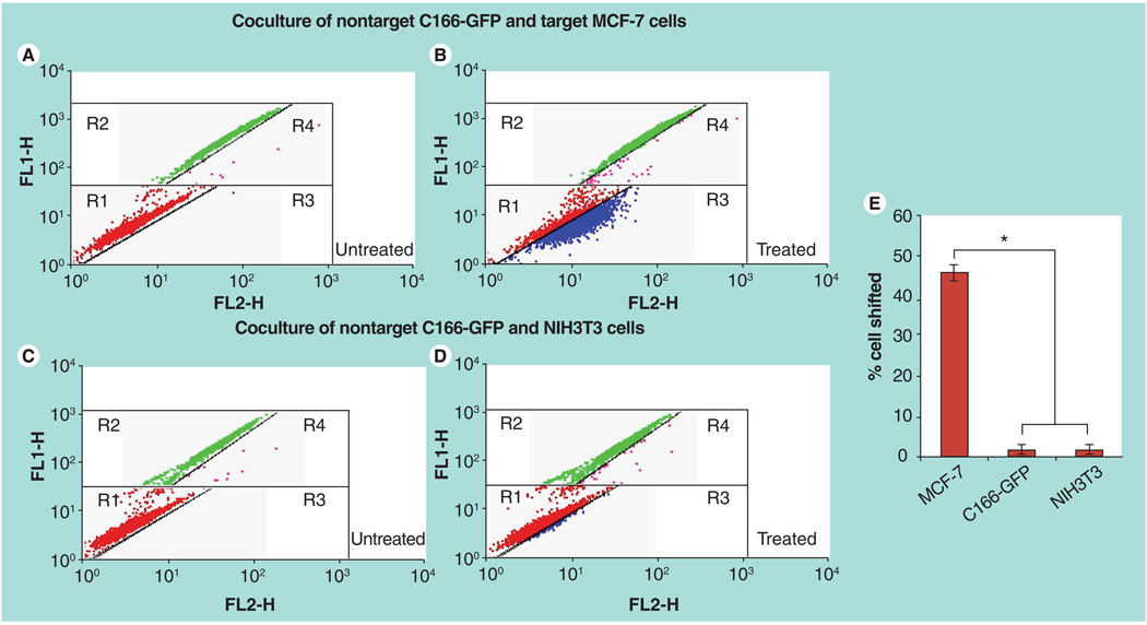
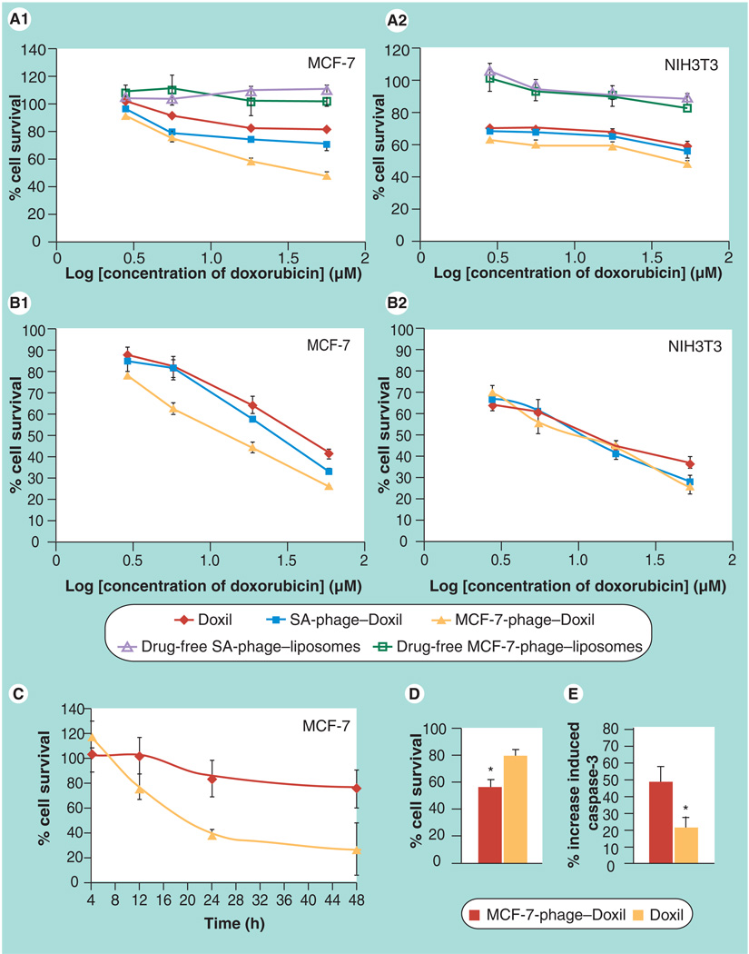
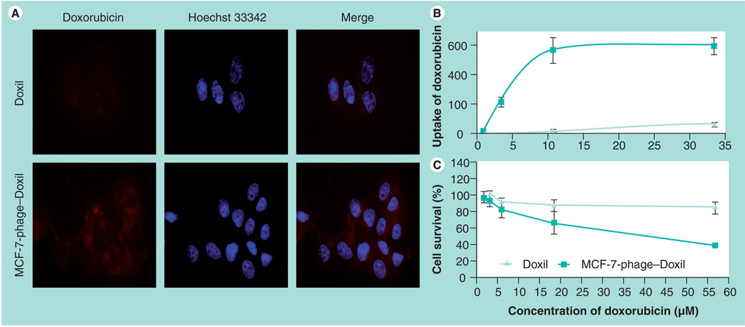
Material & methods: An 8-mer landscape library f8/8 and a biopanning protocol against MCF-7 cells were used to select a landscape phage protein bearing MCF-7-specific peptide. Size and morphology of doxorubicin-loaded liposomes modified with the tumor-specific phage fusion coat protein (phage-Doxil) were determined by dynamic light scattering and freeze-fraction electron microscopy. Topology of the phage protein in liposomes was examined by western blot. Association of phage-Doxil with MCF-7 cells was evaluated by fluorescence microscopy and fluorescence spectrometry. Selective targeting to MCF-7 was shown by FACS using a coculture model with target and nontarget cells. Phage-Doxil-induced tumor cell killing and apoptosis were confirmed by CellTiter-Blue Assay and caspase-3/CPP32 fluorometric assay.

Results: A chimeric phage fusion coat protein specific towards MCF-7 cells, identified from a phage landscape library, was directly incorporated into the liposomal bilayer of doxorubicin-loaded PEGylated liposomes (Doxil) without additional conjugation with lipophilic moieties. Western blotting confirmed the presence of both targeting peptide and pVIII coat protein in the phage-Doxil, which maintained the liposomal morphology and retained a substantial part of the incorporated drug after phage protein incorporation. The binding activity of the phage fusion pVIII coat protein was retained after incorporation into liposomes, and phage-Doxil strongly and specifically targeted MCF-7 cells, demonstrating significantly increased cytotoxicity towards target cells in vitro.

Conclusion: We present a novel and straightforward method for making tumor-targeted nanomedicines by anchoring specific phage proteins (substitute antibodies) on their surface.


Conflict of interest statement

Financial & competing interests disclosure

The authors have no other relevant affiliations or financial involvement with any organization or entity with a financial interest in or financial conflict with the subject matter or materials discussed in the manuscript apart from those disclosed.

No writing assistance was utilized in the production of this manuscript.

Figures

Figure 1
Figure 1. Production of hybrid phage fusion coat protein with genetically fused target peptide and its incorporation into liposome
Figure 2
Figure 2. Phage selectivity
Binding of the phage clone designated by the sequence of the fused foreign peptide (DMPGTVLP) with target MCF-7 cells and control MCF-10A and HepG2 cells (as percent phage recovery). The unrelated streptavidin targeting phage clone designated by the sequence of the fused foreign peptide (VPEGAFSS) was used as a control.
Figure 3
Figure 3. Characterization of Doxil® modified with MCF-7-specific phage protein (MCF-7-phage–Doxil)
(A) Western blotting pattern showing the presence of the phage protein in MCF-7-phage–Doxil. (B) The topology of the phage protein in the liposome membrane determined by the treatment with PK and terminus-specific antibodies: the N-terminus was significantly degraded in MCF-7-phage–Doxil, while the C-terminal part of the protein remained intact. (C) The liposomal morphology of MCF-7-phage–Doxil by freeze-fracture electron microscopy. (D) Size and size distribution of Doxil (D1) and MCF-7-phage–Doxil (D2). Samples were measured in triplicate and results are shown in stacked column charts. PK: Proteinase K.
Figure 4
Figure 4. FACS analysis on binding selectivity of rhodamine-labeled MCF-7-phage–liposomes in the coculture composed of nontarget C166-GFP and target MCF-7 cells and control coculture of nontarget C166-GFP and NIH3T3 cells
The dot plots were inserted into four regions (R1, R2, R3 and R4). FL1-H (green); FL2-H (red). (A) Untreated coculture of C166-GFP and MCF-7 cells. Red dots in R1 region showing the location of untreated MCF-7 cells and green dots in R2 region showing the location of untreated C166-GFP cells. (B) Coculture of C166-GFP and MCF-7 cells treated by rhodamine-labeled MCF-7-phage–liposomes. A number of MCF-7 cells (blue dots) were right shifted into R3 region, but a significantly less number of C166-GFP cells (pink dots) were right shifted into R4 region, indicating the preferential binding of MCF-7-phage–liposomes to target MCF-7 cells. (C) Untreated control coculture of C166-GFP and NIH3T3 cells. Red dots in R1 region showing the location of untreated NIH3T3 cells and green dots in R2 region showing the location of untreated C166-GFP cells. (D) Coculture of C166-GFP and NIH3T3 cells treated by rhodamine-labeled MCF-7-phage–liposomes showed the negligible binding of MCF-7-phage–liposomes to two nontargeted cells, indicating no selectivity between nontargeted NIH3T3 and C166-GFP cells. (E) Quantitative analysis of percent of cells shifted. *p < 0.05, mean ± standard deviation, n = 3
Figure 5
Figure 5. Cytotoxicity of phage–Doxil
(A) Toxicity towards MCF-7 cells (A1) and negative NIH3T3 cells (A2) after 24-h treatment with Doxil®, MCF-7-phage–Doxil, irrelevant SA-phage–Doxil, drug-free MCF-7-phage–liposomes and drug-free SA-phage–liposomes (mean ± SEM, n = 6). (B) Toxicity towards MCF-7 cells (B1) and negative NIH3T3 cells (B2) after 12-h treatment and an additional 48-h incubation in drug-free medium (mean ± SEM, n = 6). (C) Kinetics of MCF-7 cell killing in the same assay (mean ± standard deviation, n = 6). (A–C) Cytotoxicity was performed using the CellTiter-Blue® Assay. (D) Cell viability by Trypan Blue exclusion (*p < 0.05, mean ± SEM, n = 18). (E) Induction of caspase-3 in MCF-7 cells by Doxil and phage–Doxil (*p < 0.05, mean ± standard deviation, n = 3). SA-phage: Streptavidin-targeting phage.
Figure 6
Figure 6. Correlation between doxorubicin uptake and cytotoxicity of Doxil® and MCF-7-phage–Doxil
(A) Uptake of doxorubicin by MCF-7 cells was visualized by fluorescence microscopy after 30-min treatment with Doxil or MCF-7-phage–Doxil followed by 2-h incubation in drug-free medium. (B) Uptake of doxorubicin by MCF-7 cells was determined by fluorescence spectrometry after 24-h treatment with Doxil or MCF-7-phage–Doxil and was indicated as doxorubicin fluorescence intensity. (C) Cytotoxicity of Doxil and MCF-7-phage–Doxil towards MCF-7 cells after 24-h treatment (mean ± standard deviation, n = 6).

Similar articles

Cited by

References

    1. Peer D, Karp JM, Hong S, Farokhzad OC, Margalit R, Langer R. Nanocarriers as an emerging platform for cancer therapy. Nat. Nanotechnol. 2007;2:751–760. • Describes the potential of nanotechnology in revolutionizing cancer diagnosis and therapy.

    1. Alexis F, Rhee JW, Richie JP, Radovic-Moreno AF, Langer R, Farokhzad OC. New frontiers in nanotechnology for cancer treatment. Urol. Oncol. 2008;26:74–85. • Discusses recent advances of cancer nanotechnology with particular attention to nanoparticle systems that are in clinical practice or in various stages of development for cancer imaging and therapy.

    1. Sofou S. Surface-active liposomes for targeted cancer therapy. Nanomedicine (Lond.) 2007;2(5):711–724. - PubMed
    1. Noble CO, Kirpotin DB, Hayes ME, et al. Development of ligand-targeted liposomes for cancer therapy. Expert Opin. Ther. Targets. 2004;8:335–353. - PubMed
    1. Sergeeva A, Kolonin MG, Molldrem JJ, Pasqualini R, Arap W. Display technologies: application for the discovery of drug and gene delivery agents. Adv. Drug Deliv. Rev. 2006;58:1622–1654. - PMC - PubMed

Publication types