

Hybrid adipogenic implants from adipose stem cells for soft tissue reconstruction in vivo
- PMID: 20528671
- PMCID: PMC2965197
- DOI: 10.1089/ten.TEA.2010.0157
Hybrid adipogenic implants from adipose stem cells for soft tissue reconstruction in vivo
Abstract
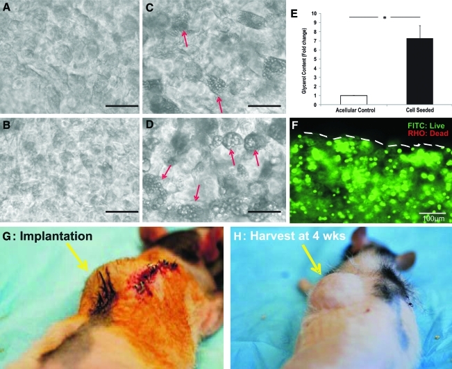
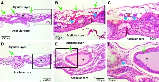
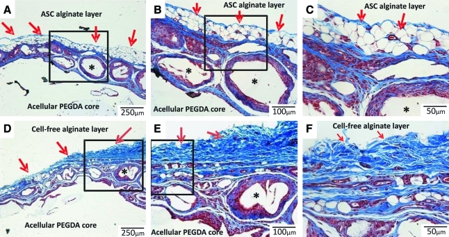
A critical barrier in tissue regeneration is scale-up. Bioengineered adipose tissue implants have been limited to ∼10 mm in diameter. Here, we devised a 40-mm hybrid implant with a cellular layer encapsulating an acellular core. Human adipose-derived stem cells (ASCs) were seeded in alginate. Poly(ethylene)glycol-diacrylate (PEGDA) was photopolymerized into 40-mm-diameter dome-shaped gel. Alginate-ASC suspension was painted onto PEGDA surface. Cultivation of hybrid constructs ex vivo in adipogenic medium for 28 days showed no delamination. Upon 4-week in vivo implantation in athymic rats, hybrid implants well integrated with host subcutaneous tissue and could only be surgically separated. Vascularized adipose tissue regenerated in the thin, painted alginate layer only if ASC-derived adipogenic cells were delivered. Contrastingly, abundant fibrous tissue filled ASC-free alginate layer encapsulating the acellular PEGDA core in control implants. Human-specific peroxisome proliferator-activated receptor-γ (PPAR-γ) was detected in human ASC-seeded implants. Interestingly, rat-specific PPAR-γ was absent in either human ASC-seeded or ASC-free implants. Glycerol content in ASC-delivered implants was significantly greater than that in ASC-free implants. Remarkably, rat-specific platelet/endothelial cell adhesion molecule (PECAM) was detected in both ASC-seeded and ASC-free implants, suggesting anastomosis of vasculature in bioengineered tissue with host blood vessels. Human nuclear staining revealed that a substantial number of adipocytes were of human origin, whereas endothelial cells of vascular wall were of chemaric human and nonhuman (rat host) origins. Together, hybrid implant appears to be a viable scale-up approach with volumetric retention attributable primarily to the acellular biomaterial core, and yet has a biologically viable cellular interface with the host. The present 40-mm soft tissue implant may serve as a biomaterial tissue expander for reconstruction of lumpectomy defects.
Figures


Similar articles
-
Comparison of readily available scaffolds for adipose tissue engineering using adipose-derived stem cells.J Plast Reconstr Aesthet Surg. 2010 May;63(5):858-64. doi: 10.1016/j.bjps.2009.01.069. Epub 2009 Apr 14. J Plast Reconstr Aesthet Surg. 2010. PMID: 19369133
-
Strategy for constructing vascularized adipose units in poly(l-glutamic acid) hydrogel porous scaffold through inducing in-situ formation of ASCs spheroids.Acta Biomater. 2017 Mar 15;51:246-257. doi: 10.1016/j.actbio.2017.01.043. Epub 2017 Jan 16. Acta Biomater. 2017. PMID: 28093366
-
Adipose-derived stem-cell-seeded non-cross-linked porcine acellular dermal matrix increases cellular infiltration, vascular infiltration, and mechanical strength of ventral hernia repairs.Tissue Eng Part A. 2015 Feb;21(3-4):475-85. doi: 10.1089/ten.TEA.2014.0235. Epub 2014 Oct 2. Tissue Eng Part A. 2015. PMID: 25156009 Free PMC article.
-
Adipose-derived stem cell differentiation as a basic tool for vascularized adipose tissue engineering.Differentiation. 2016 Jul-Aug;92(1-2):52-64. doi: 10.1016/j.diff.2016.02.003. Epub 2016 Mar 11. Differentiation. 2016. PMID: 26976717 Review.
-
Strategies for bioengineered scaffolds that support adipose stem cells in regenerative therapies.Regen Med. 2016 Sep;11(6):589-99. doi: 10.2217/rme-2016-0064. Epub 2016 Aug 3. Regen Med. 2016. PMID: 27484203 Review.
Cited by
-
Human adipose-derived stromal/stem cells induce functional CD4+CD25+FoxP3+CD127- regulatory T cells under low oxygen culture conditions.Stem Cells Dev. 2014 May 1;23(9):968-77. doi: 10.1089/scd.2013.0152. Epub 2014 Mar 11. Stem Cells Dev. 2014. PMID: 24405386 Free PMC article. Clinical Trial.
-
Enriching Nanoparticles via Acoustofluidics.ACS Nano. 2017 Jan 24;11(1):603-612. doi: 10.1021/acsnano.6b06784. Epub 2017 Jan 9. ACS Nano. 2017. PMID: 28068078 Free PMC article.
-
Epidermal growth factor-like domain protein 6 recombinant protein facilitates osteogenic differentiation in adipose stem cells via bone morphogenetic protein 2/recombinant mothers against decapentaplegic homolog 4 signaling pathway.Bioengineered. 2022 Mar;13(3):6558-6566. doi: 10.1080/21655979.2022.2037380. Bioengineered. 2022. PMID: 35220882 Free PMC article.
-
Pyrintegrin Induces Soft Tissue Formation by Transplanted or Endogenous Cells.Sci Rep. 2017 Jan 27;7:36402. doi: 10.1038/srep36402. Sci Rep. 2017. PMID: 28128224 Free PMC article.
-
Implanted adipose progenitor cells as physicochemical regulators of breast cancer.Proc Natl Acad Sci U S A. 2012 Jun 19;109(25):9786-91. doi: 10.1073/pnas.1121160109. Epub 2012 Jun 4. Proc Natl Acad Sci U S A. 2012. PMID: 22665775 Free PMC article.
References
-
- Shiffman M.A. Silicone breast implant litigation (Part 1) Med Law. 1994;13:681. - PubMed
-
- Jenkins M.E. Friedman H.I. von Recum A.F. Breast implants: facts, controversy, and speculations for future research. J Invest Surg. 1996;9:1. - PubMed
-
- Van Z.D. Heymans O. Breast implants. A review. Acta Chir Belg. 2004;104:158. - PubMed
-
- Arnez Z.M. Khan U. Pogorelec D. Planinsek F. Breast reconstruction using the free superficial inferior epigastric artery (SIEA) flap. Br J Plast Surg. 1999;52:276. - PubMed
-
- Arnez Z.M. Khan U. Pogorelec D. Planinsek F. Rational selection of flaps from the abdomen in breast reconstruction to reduce donor site morbidity. Br J Plast Surg. 1999;52:351. - PubMed
Publication types
MeSH terms
Substances
Grants and funding
LinkOut - more resources
Full Text Sources
Medical
Miscellaneous
