Clinical Trial
. 2010 Oct;85(4):290-9.
doi: 10.1111/j.1600-0609.2010.01485.x. Epub 2010 Jul 22.

First-line treatment with bortezomib rapidly stimulates both osteoblast activity and bone matrix deposition in patients with multiple myeloma, and stimulates osteoblast proliferation and differentiation in vitro

Affiliations
Free PMC article
Clinical Trial

First-line treatment with bortezomib rapidly stimulates both osteoblast activity and bone matrix deposition in patients with multiple myeloma, and stimulates osteoblast proliferation and differentiation in vitro

Thomas Lund et al. Eur J Haematol. 2010 Oct.
Free PMC article

Abstract

Objectives: The aim of the study was to investigate the effect of bortezomib on osteoblast proliferation and differentiation, as well as on bone matrix deposition for the first time in bisphosphonate-naïve, previously untreated patients with myeloma.

Methods: Twenty newly diagnosed patients received four cycles of bortezomib treatment, initially as monotherapy and then combined with a glucocorticoid from cycle two to four. Bone remodeling markers were monitored closely during treatment. Furthermore, the effects of bortezomib and a glucocorticoid on immature and mature osteoblasts were also studied in vitro.

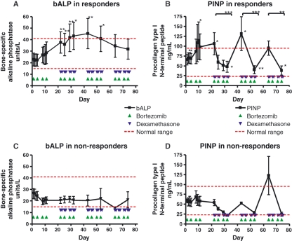
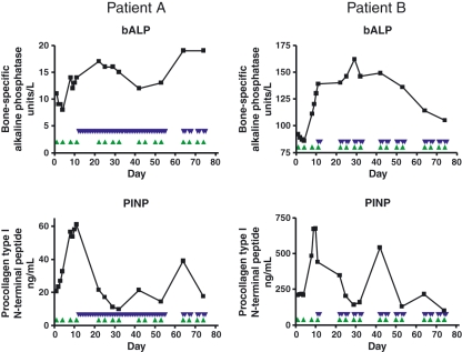
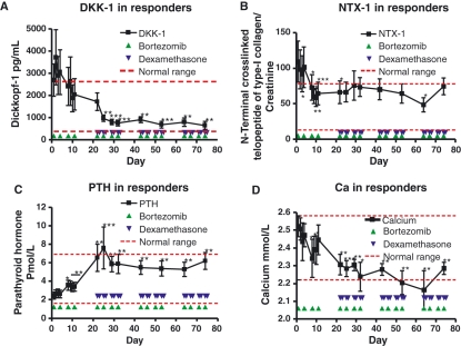
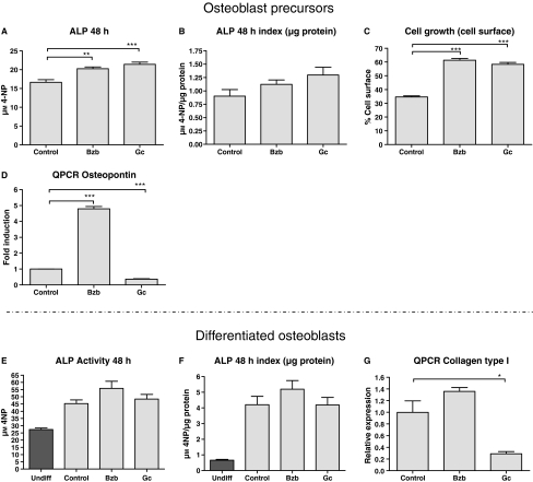
Results: Treatment with bortezomib caused a significant increase in bone-specific alkaline phosphatase and pro-collagen type I N-terminal propeptide, a novel bone formation marker. The addition of a glucocorticoid resulted in a transient decrease in collagen deposition. In vitro bortezomib induced osteoblast proliferation and differentiation. Differentiation but not proliferation was inhibited by glucocorticoid treatment.

Conclusions: Bortezomib used as first-line treatment significantly increased collagen deposition in patients with multiple myeloma and osteolytic lesions, but the addition of a glucocorticoid to the treatment transiently inhibited the positive effect of bortezomib, suggesting that bortezomib may result in better healing of osteolytic lesions when used without glucocorticoids in patients that have obtained remission with a previous therapy. The potential bone-healing properties of single-agent bortezomib are currently being explored in a clinical study in patients who have undergone high-dose therapy and autologous stem cell transplantation.


Figures

Figure 1
Figure 1
Effect of treatment according to protocol on bone-specific alkaline phosphatase (bALP) and pro-collagen Type I N-terminal peptide (PINP) in responders and non-responders. bALP increased twofold in responding patients (A). PINP showed a similar increase (B). Both values reached a maximum value at day 42. Furthermore, PINP showed a transient significant decrease every time dexamethasone was added. No significant changes were observed in non-responding patients (C–D). Response is defined as partial response or better. Results are shown as mean values ± SEM from 14 (A–B) and 4 (C–D) patients. ***P < 0.001; **P < 0.01; *P < 0.05 using a Wilcoxon signed rank test.
Figure 2
Figure 2
Effect on bone-specific alkaline phosphatase (bALP) and pro-collagen Type I N-terminal peptide (PINP) of altering the time point of administration of dexamethasone to two patients. Two patients received treatment with dexamethasone at day 12 and 11 respectively in a violation of the protocol. In both cases, bALP remained unchanged, whereas an immediate decline in PINP was observed.
Figure 3
Figure 3
Effect of treatment on Dickkopf-1 (DKK1), N-terminal crosslinked telopeptide of type I collagen (NTX-I), parathyroid hormone (PTH), and calcium in responders. DKK-1 showed a fourfold decline in responding patients (A). No changes were observed in non-responding patients (data not shown). NTX-I declined twofold, however significance is lost after the addition of dexamethasone (B). PTH increased significantly from day 8 and onward (C). The changes in PTH were mirrored by a simultaneous decrease in calcium (D). The correlation coefficient between calcium and PTH is −0.465 P < 0.0001 (data not shown). Calcium levels were corrected to albumin levels. Response is defined as partial response or better. Results are shown as mean values ± SEM from 14 patients. ***P < 0.001; **P < 0.01; *P < 0.05; using a Wilcoxon signed rank test.
Figure 4
Figure 4
Effect of bortezomib (Bzb) and prednisolone (Gc) on osteoblast precursors and differentiated osteoblasts. In pre-osteoblasts, both treatments resulted in increased levels of alkaline phosphatase (AP) (A). The significance of this was lost when adjusting for cell proliferation (i.e. total protein, B–C). Treatment with Bzb or Gc had a substantial effect on osteoblast proliferation almost doubling the area covered by cells after only 2 d (C). Osteopontin, a maker reflecting differentiation, showed a significant fivefold increase when cells were treated with Bzb; Gc on the contrary caused a decrease in osteopontin (D). No effect was observed on AP when differentiated osteoblasts were exposed to either Bzb or Gc (E–F). Treatment of differentiated osteoblasts with Gc, however, caused a fivefold decrease in collagen type I RNA, whereas collagen type I RNA remained unchanged after bortezomib exposure (G). Cells were cultured for 48 (A–C, E–F) or 24 h (D, G). Data from undifferentiated human adipose-derived stem cells (undifferentiated) are shown in E–F. Results are shown as mean values ± SEM of either five (A–C, E–F) or three replicates (D, G). ***P < 0.001; **P < 0.01; *P < 0.05; using an unpaired t-test.

Similar articles

Cited by

References

    1. Kyle RA, Gertz MA, Witzig TE, et al. Review of 1027 patients with newly diagnosed multiple myeloma. Mayo Clin Proc. 2003;78:21–33. - PubMed
    1. Farrugia AN, Atkins GJ, To LB, Pan B, Horvath N, Kostakis P, et al. Receptor activator of nuclear factor-kappaB ligand expression by human myeloma cells mediates osteoclast formation in vitro and correlates with bone destruction in vivo. Cancer Res. 2003;63:5438–45. - PubMed
    1. Giuliani N, Bataille R, Mancini C, Lazzaretti M, Barille S. Myeloma cells induce imbalance in the osteoprotegerin/osteoprotegerin ligand system in the human bone marrow environment. Blood. 2001;98:3527–33. - PubMed
    1. Michigami T, Shimizu N, Williams PJ, Niewolna M, Dallas SL, Mundy GR, et al. Cell-cell contact between marrow stromal cells and myeloma cells via VCAM-1 and alpha(4)beta(1)-integrin enhances production of osteoclast-stimulating activity. Blood. 2000;96:1953–60. - PubMed
    1. Pearse RN, Sordillo EM, Yaccoby S, Wong BR, Liau DF, Colman N, et al. Multiple myeloma disrupts the TRANCE/osteoprotegerin cytokine axis to trigger bone destruction and promote tumor progression. Proc Natl Acad Sci U S A. 2001;98:11581–6. - PMC - PubMed

Publication types

MeSH terms