Review
. 2010 Nov;34(6):1076-112.
doi: 10.1111/j.1574-6976.2010.00231.x.

RTX proteins: a highly diverse family secreted by a common mechanism

Affiliations
Free PMC article
Review

RTX proteins: a highly diverse family secreted by a common mechanism

Irena Linhartová et al. FEMS Microbiol Rev. 2010 Nov.
Free PMC article

Erratum in

Abstract

Repeats-in-toxin (RTX) exoproteins of Gram-negative bacteria form a steadily growing family of proteins with diverse biological functions. Their common feature is the unique mode of export across the bacterial envelope via the type I secretion system and the characteristic, typically nonapeptide, glycine- and aspartate-rich repeats binding Ca(2+) ions. In this review, we summarize the current state of knowledge on the organization of rtx loci and on the biological and biochemical activities of therein encoded proteins. Applying several types of bioinformatic screens on the steadily growing set of sequenced bacterial genomes, over 1000 RTX family members were detected, with the biological functions of most of them remaining to be characterized. Activities of the so far characterized RTX family members are then discussed and classified according to functional categories, ranging from the historically first characterized pore-forming RTX leukotoxins, through the large multifunctional enzymatic toxins, bacteriocins, nodulation proteins, surface layer proteins, up to secreted hydrolytic enzymes exhibiting metalloprotease or lipase activities of industrial interest.


Figures
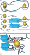
Fig. 1
Fig. 1
The parallel β-roll structure of the RTX repeats. Top view (a) and side view (b) projection of the RTX repeats comprising residues Gly333 and Phe376 from the three-dimensional model of alkaline protease of Pseudomonas aeruginosa (Baumann et al., 1993). The protein backbone, short β-strands and Ca2+ ions are represented as grey ribbon, cyan arrows and yellow balls, respectively. (c) A detailed view of the calcium-binding site within the RTX repeats. Each nonapeptide motif forms two half-sites for Ca2+ binding, where each Ca2+ ion is bound in a six coordinate site between two consecutive turns. The first turn contributes the main chain carbonyls of Asn343 and Val345, and one carboxyl oxygen of Asn347. The second turn contributes the carbonyls of Gly360 and Leu362, as well as the carboxyl oxygen of Asp365. Residues whose side chains directly coordinate the Ca2+ ion are highlighted. The carbon atoms of the side chains are green, nitrogens are blue, oxygens are red.
Fig. 2
Fig. 2
The schematic depiction of the TISS assembly operation. Upon recognition of a C-terminal secretion signal on a given RTX protein translocation substrate, the inner membrane complex formed by an energy-providing ABC transporter and a MFP contacts the trimeric OMP. A sealed channel–tunnel assembly spanning across the entire Gram-negative bacterial cell envelope is formed, through which the RTX protein is exported in a single step from the bacterial cytoplasm directly to the external bacterial surface, without transiting through the periplasmic space. While concentrations of Ca2+ ions are typically <100 nM in bacterial cytoplasm, allowing for maintenance of an unfolded RTX domain, millimolar calcium concentrations are typically encountered in host extracellular space colonized by pathogenic bacteria. Loading of RTX repeats of the secreted protein by Ca2+ ions then promotes its folding and acquisition of biological activity.
Fig. 3
Fig. 3
(a) The schematic representation of the rtx gene clusters of Escherichia coli, Pasteurella haemolytica, Bordetella pertussis and Vibrio cholerae. The arrows represent coding regions and transcriptional directions of the rtx genes deposited under the following GenBank accession numbers: E. coli (NC 000913); P. haemolytica PHL213 (NZ AASA00000000); B. pertussis Tohama I (NC 002929) and V. cholerae N16961 (NC 002505). (b) Domain structures of the RTX cytotoxins HlyA (E. coli), LktA (P. haemolytica), CyaA (B. pertussis) and MARTXVc (V. cholerae) with indication of sites of post-translational modification of internal lysines by covalent attachment of fatty acyl residues. The functional domain labelling is as follows: H, haemolytic domain; RTX, RTX domain; AC, adenylate cyclase domain; ACD, actin cross-linking domain; RID, Rho inactivation domain; CPD, cystein protease domain.
Fig. 4
Fig. 4
The schematic representation of rtx protease gene clusters. The arrows represent coding regions and transcriptional directions of the genes deposited under the GenBank accession numbers: Pseudomonas aeruginosa (AY003006, X64558), Photorhabdus (AY230750), Pseudomonas fluorescens SIK W1 (AF083061), P. fluorescens B52 (AF216700, AF216701, AF216702), Pseudomonas brassicacearum (AF286062), Erwinia amylovora (Y19002), Erwinia chrysanthemi (M60395), Proteus mirabilis (AF064762).
Fig. 5
Fig. 5
The schematic representation of rtx lipase gene clusters of lipases. The arrows represent coding regions and transcriptional directions of the genes deposited under the GenBank accession numbers: Serratia marcescens (D49826), Pseudomonas fluorescens SIK W1 (AF083061), P. fluorescens no. 33 (AB015053), Pseudomonas brassicacearum (AF286062).
Fig. 6
Fig. 6
Comparison of size and pI distribution of the bulk of bacterial proteins and of the sum of characterized and putative RTX proteins. Molecular weight (MW) and pI was calculated for the bulk of bacterial proteins (about 2.75 mil., shown in black) and for the identified and predicted RTX proteins (1024 proteins, shown as red crosses) using the program pepstat, a part of the emboss package (Rice et al., 2000). MW was log base 10 transformed (y-axis) and plotted against calculated pI (x-axis) in statistical package r version 2.9.0 (http://www.r-project.org/).
Fig. 7
Fig. 7
Use of a simple RTX building unit in generation of complex biological functionalities. By maintaining an unfolded state which allows a post-translational secretion from the calcium-depleted cytoplasm, and by promoting protein folding upon the binding of calcium ions (yellow balls) in the extracellular environment, the C-terminal assemblies of glycine- and aspartate-rich nonapeptide RTX repeat units first assist in the translocation of even very large RTX proteins. These transit across the entire Gram-negative bacterial cell envelope in a single step mediated by the dedicated the type I secretion machinery recognizing unprocessed C-terminal secretion signals. Proteins using this secretion pathway exhibit a very broad range of biological functions in colonizing diverse host environments. RTX proteins were found to exert activities like structural proteins involved in protective S-layer formation and motility of bacteria, in colonization of root nodules of plants by symbiotic bacteria, serving as bacteriocins on other bacteria, exerting hydrolase activities, or playing a prominent role as essential colonization and virulence factors of bacteria in animal hosts, respectively. Besides of a large group of pore-forming leukotoxins a particular sophistication of function is observed for the very large (thousands of residues long) RTX toxins consisting of multiple domains exhibiting enzymatic and cytotoxic activities (MARTX).

References

    1. Adusu TE, Conlon PD, Shewen PE, Black WD. Pasteurella haemolytica leukotoxin induces histamine release from bovine pulmonary mast cells. Can J Vet Res. 1994;58:1–5. - PMC - PubMed
    1. Ahn JH, Pan JG, Rhee JS. Identification of the tliDEF ABC transporter specific for lipase in Pseudomonas fluorescens SIK W1. J Bacteriol. 1999;181:1847–1852. - PMC - PubMed
    1. Akatsuka H, Kawai E, Omori K, Komatsubara S, Shibatani T, Tosa T. The lipA gene of Serratia marcescens which encodes an extracellular lipase having no N-terminal signal peptide. J Bacteriol. 1994;176:1949–1956. - PMC - PubMed
    1. Akatsuka H, Kawai E, Omori K, Shibatani T. The three genes lipB, lipC, and lipD involved in the extracellular secretion of the Serratia marcescens lipase which lacks an N-terminal signal peptide. J Bacteriol. 1995;177:6381–6389. - PMC - PubMed
    1. Altschul SF, Madden TL, Schaffer AA, Zhang J, Zhang Z, Miller W, Lipman DJ. Gapped BLAST and PSI-BLAST: a new generation of protein database search programs. Nucleic Acids Res. 1997;25:3389–3402. - PMC - PubMed

Publication types

MeSH terms