Malignant fibrous histiocytoma of the distal femur after an arthroscopic anterior cruciate ligament reconstruction: A case report and a review of the literature
- PMID: 20529315
- PMCID: PMC2889898
- DOI: 10.1186/1471-2407-10-264
Malignant fibrous histiocytoma of the distal femur after an arthroscopic anterior cruciate ligament reconstruction: A case report and a review of the literature
Abstract
Background: Malignant degeneration in association with orthopaedic implants is a known but rare complication. To our knowledge, no case of osseous malignant fibrous histiocytoma after anterior cruciate ligament reconstruction is reported in the literature.
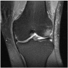
Case presentation: We report a 29-year-old male Turkish patient who presented with severe pain in the operated knee joint 40 months after arthroscopic anterior cruciate ligament reconstruction. X-ray and MR imaging showed a large destructive tumor in the medial femoral condyle. Biopsy determined a malignant fibrous histiocytoma. After neoadjuvant chemotherapy, wide tumor resection and distal femur reconstruction with a silver-coated non-cemented tumor knee joint prosthesis was performed. Adjuvant chemotherapy was continued according to the EURAMOS 1 protocol.
Conclusions: Though secondary malignant degeneration after orthopaedic implants or prostheses is not very likely, the attending physician should take this into consideration, especially if symptoms worsen severely over a short period of time.
Figures


Similar articles
-
Pleomorphic malignant fibrous histiocytoma at the site of an arthroscopic reconstruction of the anterior cruciate ligament. A case report.J Bone Joint Surg Am. 2005 Feb;87(2):404-9. doi: 10.2106/JBJS.C.01207. J Bone Joint Surg Am. 2005. PMID: 15687167 No abstract available.
-
Hamstring insertion site healing after anterior cruciate ligament reconstruction in patients with symptomatic hardware or repeat rupture: a histologic study in 12 patients.Arthroscopy. 2003 Nov;19(9):948-54. doi: 10.1016/j.arthro.2003.09.007. Arthroscopy. 2003. PMID: 14608313
-
Variations in Knee Kinematics After ACL Injury and After Reconstruction Are Correlated With Bone Shape Differences.Clin Orthop Relat Res. 2017 Oct;475(10):2427-2435. doi: 10.1007/s11999-017-5368-8. Clin Orthop Relat Res. 2017. PMID: 28451863 Free PMC article.
-
[Combined posterior and anterior cruciate ligament reconstruction : Arthroscopic treatment with the GraftLink® system].Oper Orthop Traumatol. 2019 Feb;31(1):20-35. doi: 10.1007/s00064-018-0580-6. Epub 2018 Dec 18. Oper Orthop Traumatol. 2019. PMID: 30564843 Review. German.
-
Fungal osteomyelitis after arthroscopic anterior cruciate ligament reconstruction: a case report with review of the literature.Knee. 2012 Oct;19(5):728-31. doi: 10.1016/j.knee.2011.10.007. Epub 2011 Dec 30. Knee. 2012. PMID: 22209694 Review.
Cited by
-
Successful treatment of advanced malignant fibrous histiocytoma of the right forearm with apatinib: a case report.Onco Targets Ther. 2016 Feb 5;9:643-7. doi: 10.2147/OTT.S96133. eCollection 2016. Onco Targets Ther. 2016. PMID: 26917971 Free PMC article.
-
Pain and fracture after anterior cruciate ligament reconstruction caused by giant cell tumour of the distal femur.BMJ Case Rep. 2013 Sep 26;2013:bcr2013010422. doi: 10.1136/bcr-2013-010422. BMJ Case Rep. 2013. PMID: 24072827 Free PMC article.
-
Synovium as a widespread pathway to the adjacent joint in undifferentiated high-grade pleomorphic sarcoma of the tibia: A case report.Medicine (Baltimore). 2018 Feb;97(8):e9870. doi: 10.1097/MD.0000000000009870. Medicine (Baltimore). 2018. PMID: 29465573 Free PMC article.
-
A novel TMTC2-NTRK3 fusion in undifferentiated high-grade pleomorphic sarcoma.J Cancer Res Clin Oncol. 2022 Oct;148(10):2933-2937. doi: 10.1007/s00432-022-04249-x. Epub 2022 Aug 7. J Cancer Res Clin Oncol. 2022. PMID: 35933643 Free PMC article.
References
Publication types
MeSH terms
LinkOut - more resources
Full Text Sources

