. 2010 Jun 8:10:14.
doi: 10.1186/1472-6807-10-14.

Active site plasticity revealed from the structure of the enterobacterial N-ribohydrolase RihA bound to a competitive inhibitor

Affiliations

Active site plasticity revealed from the structure of the enterobacterial N-ribohydrolase RihA bound to a competitive inhibitor

Gianpiero Garau et al. BMC Struct Biol. .

Abstract

Background: Pyrimidine-preferring N-ribohydrolases (CU-NHs) are a class of Ca2+-dependent enzymes that catalyze the hydrolytic cleavage of the N-glycosidic bond in pyrimidine nucleosides. With the exception of few selected organisms, their physiological relevance in prokaryotes and eukaryotes is yet under investigation.

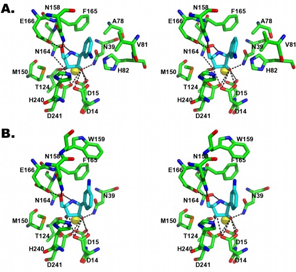
Results: Here, we report the first crystal structure of a CU-NH bound to a competitive inhibitor, the complex between the Escherichia coli enzyme RihA bound to 3, 4-diaminophenyl-iminoribitol (DAPIR) to a resolution of 2.1 A. The ligand can bind at the active site in two distinct orientations, and the stabilization of two flexible active site regions is pivotal to establish the interactions required for substrate discrimination and catalysis.

Conclusions: A comparison with the product-bound RihA structure allows a rationalization of the structural rearrangements required for an enzymatic catalytic cycle, highlighting a substrate-assisted cooperative motion, and suggesting a yet overlooked role of the conserved His82 residue in modulating product release. Differences in the structural features of the active sites in the two homologous CU-NHs RihA and RihB from E. coli provide a rationale for their fine differences in substrate specificity. These new findings hint at a possible role of CU-NHs in the breakdown of modified nucleosides derived from RNA molecules.


Figures

Figure 1
Figure 1
Ligands of the E. coli pyrimidine-preferring NH RihA. A) Uridine and B) cytidine, the natural substrates. C) The competitive inhibitor DAPIR. The iminoribitol moiety resembles the features of the transition state of the hydrolytic reaction, including a partial double bond character of the C1'-O4' bond, an elongated N-glycosidic bond, and a partial positive charge at the sugar ring.
Figure 2
Figure 2
Crystal structure of RihA bound to DAPIR. A) The RihA monomer, with α-helices coloured brown, β-strands blue, and loop regions in cyan. The active site Ca2+ ion is shown as a yellow sphere, and the DAPIR molecule as sticks coloured according to atom type. B) The RihA tetramer, as observed in the asymmetric unit of the RihA-DAPIR complex crystals.
Figure 3
Figure 3
Structural features of the RihA-DAPIR complex. A) Superposition of the four crystallographically-independent molecules of RihA. The Cα trace of the four RihA monomers are shown in grey, and the αL (red) and α9 (green) segments are labelled for clarity. The four independent RihA chains are virtually undistinguishable. The αL loop is completely visible in chain A, while the full α9 helix could be traced only in chain D, as per nomenclature of the deposited coordinates. B) The DAPIR molecule bound to the active site of chain A superimposed with a (2mFo-DFc, ϕc) shake-omit electron density map contoured at 1.2σ. C) Octacoordination of the active site Ca2+ ion. Five oxygen atoms from RihA amino acid residues, the O2' and O3' hydroxyls of DAPIR, and an highly-ordered water molecule complete the coordination sphere of the metal.
Figure 4
Figure 4
Dual mode of DAPIR binding to RihA. A) Stereo view of the active site of molecule A, showing the closed αL segment with the inhibitor N3 group pointing towards helix α9. B) Stereo view of the active site of molecule B, where the αL segment is flexible and the N3 amino group of DAPIR points in the opposite direction compared to panel C.
Figure 5
Figure 5
A comparison of the structures of the pyrimidine-preferring CU-NHs RihA and RihB from E. coli. A) Superposition of the RihA and RihB monomers, shown as Cα traces. The α9 helix conformation differs significantly between the two proteins and is indicative of the plasticity of the flexible regions in NHs to adapt to different ligands. B) Model of interactions between RihA and uridine (top) or cytidine (bottom).
Figure 6
Figure 6
Active site versatility of RihA. Molecular surfaces at the RihA active sites of two independent molecules. Two orthogonal views are shown, highlighting the existence of empty cavities that may accommodate different types of substrates. Panels A) and B) depict the surfaces of chain A and chain D, respectively.

Similar articles

Cited by

References

    1. Schramm VL. Enzymatic N-riboside scission in RNA and RNA precursors. Curr Opin Chem Biol. 1997;1(4):589–590. doi: 10.1016/S1367-5931(97)80056-7. - DOI - PubMed
    1. Versees W, Steyaert J. Catalysis by nucleoside hydrolases. Curr Opin Struct Biol. 2003;13(6):731–738. doi: 10.1016/j.sbi.2003.10.002. - DOI - PubMed
    1. Petersen C, Moller LB. The RihA, RihB, and RihC ribonucleoside hydrolases of Escherichia coli. Substrate specificity, gene expression, and regulation. J Biol Chem. 2001;276(2):884–894. doi: 10.1074/jbc.M008300200. - DOI - PubMed
    1. Kurtz JE, Exinger F, Erbs P, Jund R. The URH1 uridine ribohydrolase of Saccharomyces cerevisiae. Curr Genet. 2002;41(3):132–141. doi: 10.1007/s00294-002-0296-9. - DOI - PubMed
    1. Liang L, He X, Liu G, Tan H. The role of a purine-specific nucleoside hydrolase in spore germination of Bacillus thuringiensis. Microbiology. 2008;154(Pt 5):1333–1340. doi: 10.1099/mic.0.2007/014399-0. - DOI - PubMed

Publication types

MeSH terms

LinkOut - more resources