. 2010 Jun 8:10:265.
doi: 10.1186/1471-2407-10-265.

Methylthioadenosine (MTA) inhibits melanoma cell proliferation and in vivo tumor growth

Affiliations

Methylthioadenosine (MTA) inhibits melanoma cell proliferation and in vivo tumor growth

Pedro Andreu-Pérez et al. BMC Cancer. .

Abstract

Background: Melanoma is the most deadly form of skin cancer without effective treatment. Methylthioadenosine (MTA) is a naturally occurring nucleoside with differential effects on normal and transformed cells. MTA has been widely demonstrated to promote anti-proliferative and pro-apoptotic responses in different cell types. In this study we have assessed the therapeutic potential of MTA in melanoma treatment.

Methods: To investigate the therapeutic potential of MTA we performed in vitro proliferation and viability assays using six different mouse and human melanoma cell lines wild type for RAS and BRAF or harboring different mutations in RAS pathway. We also have tested its therapeutic capabilities in vivo in a xenograft mouse melanoma model and using variety of molecular techniques and tissue culture we investigated its anti-proliferative and pro-apoptotic properties.

Results: In vitro experiments showed that MTA treatment inhibited melanoma cell proliferation and viability in a dose dependent manner, where BRAF mutant melanoma cell lines appear to be more sensitive. Importantly, MTA was effective inhibiting in vivo tumor growth. The molecular analysis of tumor samples and in vitro experiments indicated that MTA induces cytostatic rather than pro-apoptotic effects inhibiting the phosphorylation of Akt and S6 ribosomal protein and inducing the down-regulation of cyclin D1.

Conclusions: MTA inhibits melanoma cell proliferation and in vivo tumor growth particularly in BRAF mutant melanoma cells. These data reveal a naturally occurring drug potentially useful for melanoma treatment.


Figures
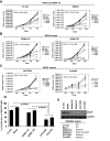
Figure 1
Figure 1
Methylthioadenosine inhibits melanoma cell growth. Proliferation assays of (A) 37-31E and MeWo (wild type RAS BRAF), (B) SKMel147 and SKMel103 (NRASQ61L mutated) and (C) UACC903 and Colo829 (BRAFV600E mutated) melanoma cells in complete medium with increasing concentrations of Methylthioadenosine (MTA). Medium was changed adding fresh MTA every 48 h. Proliferation assays were performed by counting the number of viable cells using Guava-Viacount reagent (Guava Technologies) in a cell counter (Viacount) at the time points indicated. Asterisk indicate p < 0.05. (D) Graph showing the percentage of cell proliferation inhibition at 96 h treated with 10 μM MTA. p-values were calculated performing a t-student test. (E) Western-blot showing the levels of MTAP in the different cell lines. The mutational status of CDK2NA for the different cell lines is showed in the table.
Figure 2
Figure 2
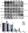
Methylthioadenosine (MTA) decreases melanoma cell viability. Clonogenic assays of 37-31E, MeWo, SKMel147, SKMel103, UACC903 and Colo 829 melanoma cells in complete medium with increasing concentrations of Methylthioadenosine (MTA). Three hundred cells were seeded clones were visualized by crystal violet staining and counted. Fresh media was changed every 48 h. Graphs represent the percentage reduction in the number of clones upon different treatments. p-value was calculated performing a Wilcoxon-signed rank test (Vassar Stats).
Figure 3
Figure 3
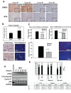
Methylthioadenosine (MTA) inhibits in vivo tumor growth and decreases the activation of RAS and PI3K tumor signaling pathways. (A) 37-31E melanoma cell line was subcutaneously injected into immunocompetent FVB/N mice. The animal groups were treated with DMSO (Control) (n = 7) or methylthioadenosine (MTA) (n = 7). Tumor size was calculated as described in materials and methods. p-values were calculated performing a t-student test. Representative photographs of three paired untreated and MTA-treated excised melanoma tumors are showed on the right. (B) Immunostaining of paraffin-embedded tumor sections showing the levels of p-Erk1/2, p-Akt and p-S6 (20×). Insets show a 40× detail of the picture. Representative images from two different untreated and treated tumors are shown.
Figure 4
Figure 4
Methylthioadenosine (MTA) promotes cytostatic effects rather than pro-apoptotic effects in melanoma tumors and cells. (A) Tumor sections from mice treated either with DMSO or MTA were stained with cyclin D1 and Ki67 antibodies. Representative pictures are shown. (B) Quantification of apoptotic cells within the tumors. Paraffin-embedded tumor samples were subjected to TUNEL assay or stained against cleaved caspase-3. Graph shows the quantification of the TUNEL assay. Positive cells from ten fields (20×) per sample were quantified and the average number of cells per field was calculated. (C) Upper graph, quantification by qRT-PCR of VEGF levels in tumor samples. Lower graph, qRT-PCR of VEGF expression levels. 37-31E were untreated (Control) or treated for 48 h with 10 μM of MTA in complete medium. Microvessel's density quantification in xenografts. Graph shows CD31 fluorescence per μm2. Representative pictures are showed on the right. p-values were calculated performing a t-student test. (D) 37-31E cells were treated with MTA (10 μM) for the time points indicated. Fifty micrograms of total lysates were resolved by PAGE-SDS. p-Bad, cleaved-caspase3, p-S6 and cyclin D1 protein levels are showed. GAPDH is used as a loading control. (E) MTA treatment induces a slowdown cell cycle G1 phase. Cells were grown in complete medium for 48 h in the presence or absence of MTA (10 μM). Cell cycle analysis was measured in triplicates using Cell Cycle Analysis Guava-Viacount reagent (Guava Technologies). Average of the three samples in each phase of the cell cycle are shown. p-values were calculated performing a t-student test (* = p < 0.05; ** = p < 0.01).

Similar articles

Cited by

References

    1. Savarese TM, Ghoda LY, Dexter DL, Parks RE Jr. Conversion of 5'-deoxy-5'-methylthioadenosine and 5'-deoxy-5'-methylthioinosine to methionine in cultured human leukemic cells. Cancer Res. 1983;43(10):4699–4702. - PubMed
    1. Olopade OI, Pomykala HM, Hagos F, Sveen LW, Espinosa R, Dreyling MH, Gursky S, Stadler WM, Le Beau MM, Bohlander SK. Construction of a 2.8-megabase yeast artificial chromosome contig and cloning of the human methylthioadenosine phosphorylase gene from the tumor suppressor region on 9p21. Proc Natl Acad Sci USA. 1995;92(14):6489–6493. doi: 10.1073/pnas.92.14.6489. - DOI - PMC - PubMed
    1. Chin L, Garraway LA, Fisher DE. Malignant melanoma: genetics and therapeutics in the genomic era. Genes Dev. 2006;20(16):2149–2182. doi: 10.1101/gad.1437206. - DOI - PubMed
    1. Bishop DT, Demenais F, Iles MM, Harland M, Taylor JC, Corda E, Randerson-Moor J, Aitken JF, Avril MF, Azizi E. Genome-wide association study identifies three loci associated with melanoma risk. Nat Genet. 2009;41(8):920–925. doi: 10.1038/ng.411. - DOI - PMC - PubMed
    1. Stevens AP, Spangler B, Wallner S, Kreutz M, Dettmer K, Oefner PJ, Bosserhoff AK. Direct and tumor microenvironment mediated influences of 5'-deoxy-5'-(methylthio)adenosine on tumor progression of malignant melanoma. J Cell Biochem. 2009;106(2):210–219. doi: 10.1002/jcb.21984. - DOI - PubMed

Publication types

MeSH terms