

Heme-heme and heme-ligand interactions in the di-heme oxygen-reducing site of cytochrome bd from Escherichia coli revealed by nanosecond absorption spectroscopy
- PMID: 20529691
- PMCID: PMC3990236
- DOI: 10.1016/j.bbabio.2010.05.010
Heme-heme and heme-ligand interactions in the di-heme oxygen-reducing site of cytochrome bd from Escherichia coli revealed by nanosecond absorption spectroscopy
Abstract
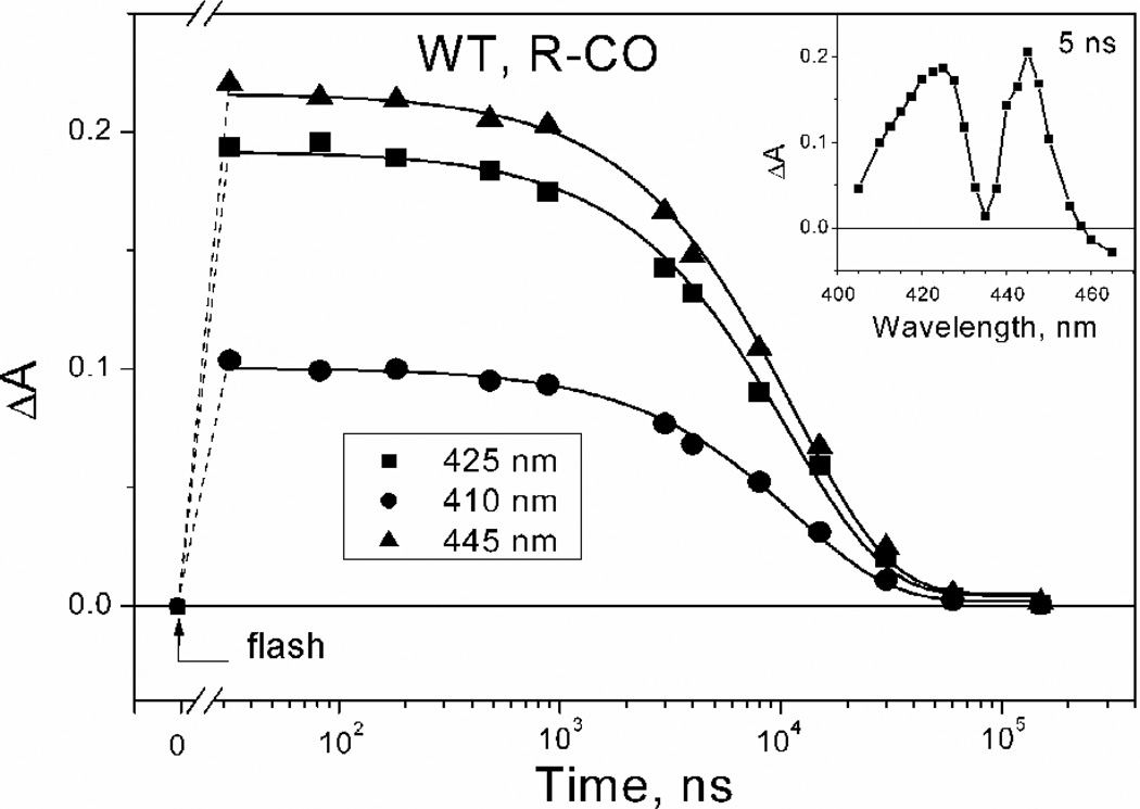
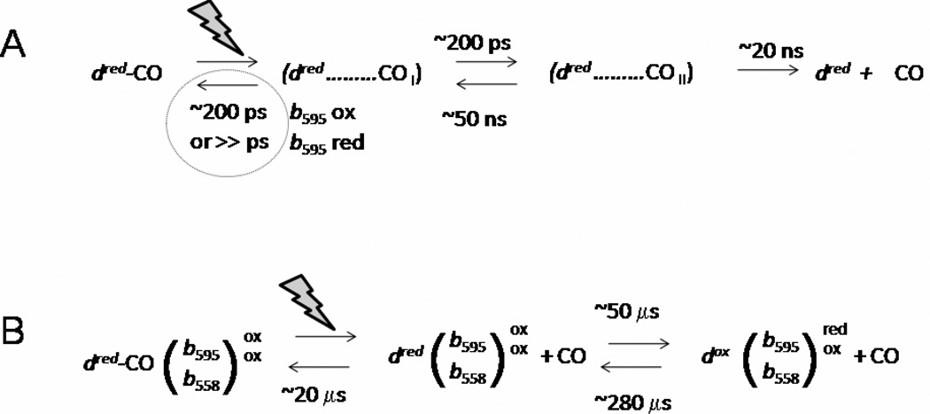
Cytochrome bd is a terminal quinol:O(2) oxidoreductase of respiratory chains of many bacteria. It contains three hemes, b(558), b(595), and d. The role of heme b(595) remains obscure. A CO photolysis/recombination study of the membranes of Escherichia coli containing either wild type cytochrome bd or inactive E445A mutant was performed using nanosecond absorption spectroscopy. We compared photoinduced changes of heme d-CO complex in one-electron-reduced, two-electron-reduced, and fully reduced states of cytochromes bd. The line shape of spectra of photodissociation of one-electron-reduced and two-electron-reduced enzymes is strikingly different from that of the fully reduced enzyme. The difference demonstrates that in the fully reduced enzyme photolysis of CO from heme d perturbs ferrous heme b(595) causing loss of an absorption band centered at 435 nm, thus supporting interactions between heme b(595) and heme d in the di-heme oxygen-reducing site, in agreement with previous works. Photolyzed CO recombines with the fully reduced enzyme monoexponentially with tau approximately 12 micros, whereas recombination of CO with one-electron-reduced cytochrome bd shows three kinetic phases, with tau approximately 14 ns, 14 micros, and 280 micros. The spectra of the absorption changes associated with these components are different in line shape. The 14 ns phase, absent in the fully reduced enzyme, reflects geminate recombination of CO with part of heme d. The 14-micros component reflects bimolecular recombination of CO with heme d and electron backflow from heme d to hemes b in approximately 4% of the enzyme population. The final, 280-micros component, reflects return of the electron from hemes b to heme d and bimolecular recombination of CO in that population. The fact that even in the two-electron-reduced enzyme, a nanosecond geminate recombination is observed, suggests that namely the redox state of heme b(595), and not that of heme b(558), controls the pathway(s) by which CO migrates between heme d and the medium.
2010 Elsevier B.V. All rights reserved.
Figures


References
-
- Poole RK, Cook GM. Redundancy of aerobic respiratory chains in bacteria? Routes, reasons and regulation. Adv. Microb. Physiol. 2000;43:165–224. - PubMed
-
- Junemann S. Cytochrome bd terminal oxidase. Biochim. Biophys. Acta. 1997;1321:107–127. - PubMed
-
- Borisov VB. Cytochrome bd: structure and properties. Biochemistry (Moscow) 1996;61:565–574. (translated from Biokhimiya (in Russian) (1996), 61, 786–799). - PubMed
-
- Tsubaki M, Hori H, Mogi T. Probing molecular structure of dioxygen reduction site of bacterial quinol oxidases through ligand binding to the redox metal centers. J. Inorg. Biochem. 2000;82:19–25. - PubMed
-
- Borisov VB, Verkhovsky MI. Oxygen as acceptor [Chapter 3.2.7] In: Böck A, Curtiss R III, Kaper JB, Neidhardt FC, Nyström T, Rudd KE, Squires CL, editors. EcoSal - Escherichia coli and Salmonella: cellular and molecular biology. Washington, DC: ASM Press; 2009. < http://www.ecosal.org>.
Publication types
MeSH terms
Substances
Grants and funding
LinkOut - more resources
Full Text Sources
Molecular Biology Databases
Miscellaneous
