. 2010 Oct;29(10):1739-58.
doi: 10.1109/TMI.2010.2051680. Epub 2010 Jun 7.

Robust super-resolution volume reconstruction from slice acquisitions: application to fetal brain MRI

Affiliations

Robust super-resolution volume reconstruction from slice acquisitions: application to fetal brain MRI

Ali Gholipour et al. IEEE Trans Med Imaging. 2010 Oct.

Abstract

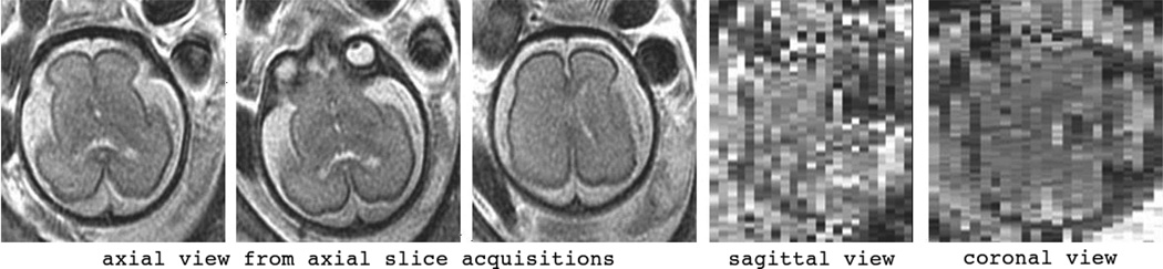
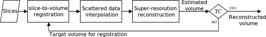
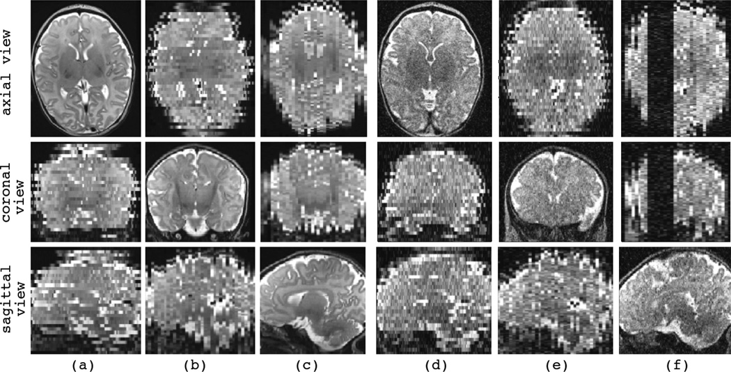
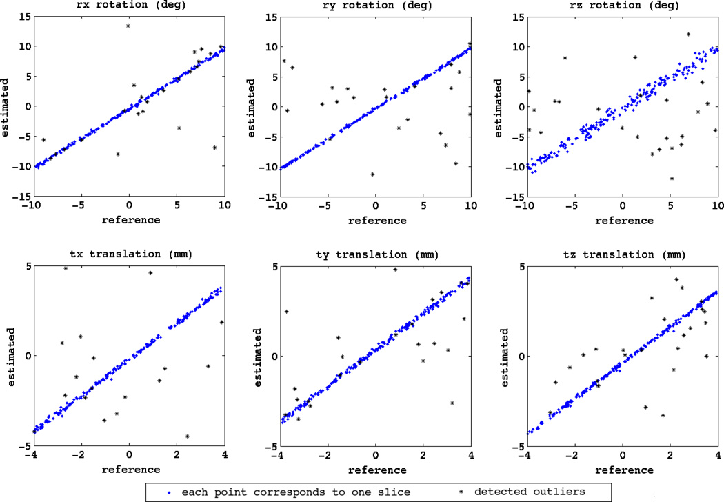
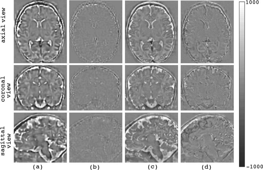
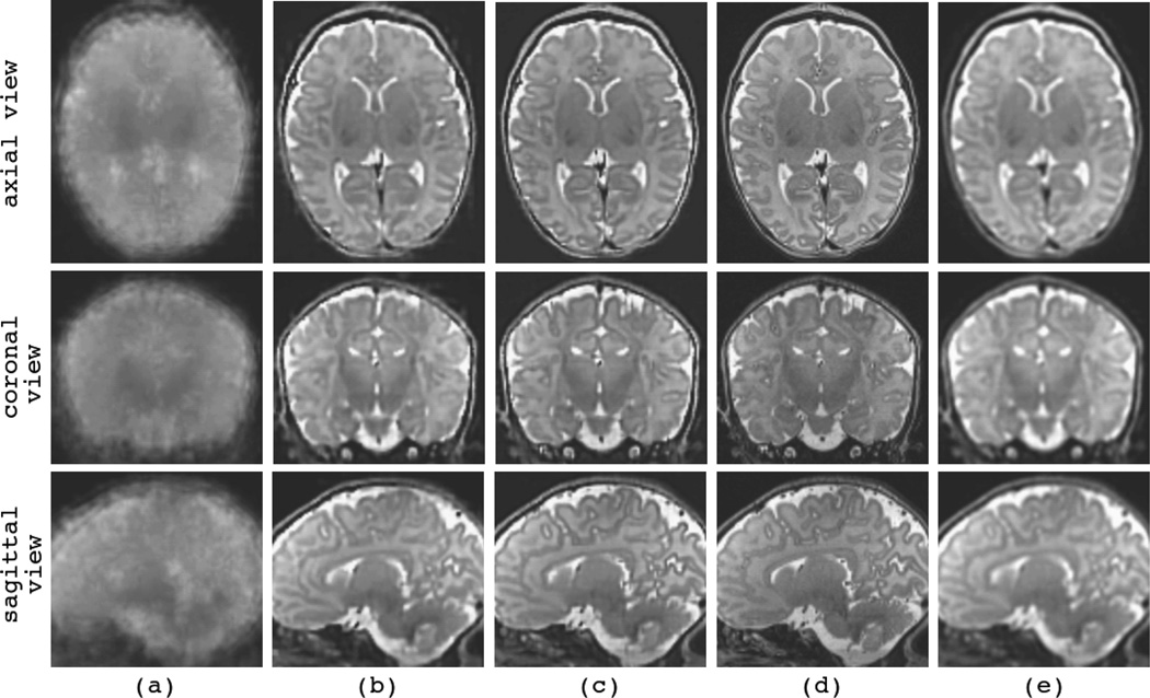
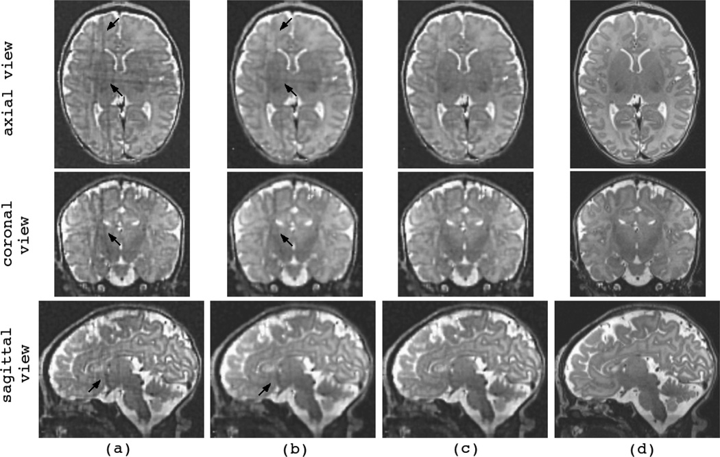
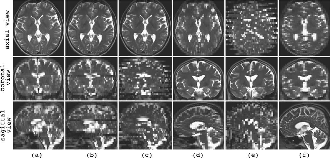
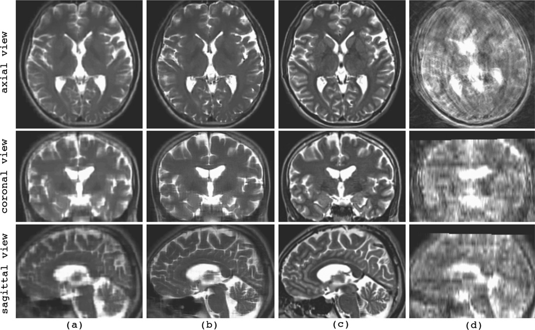
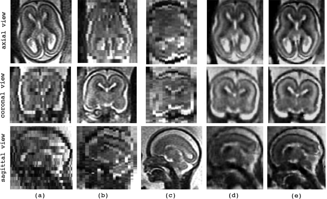
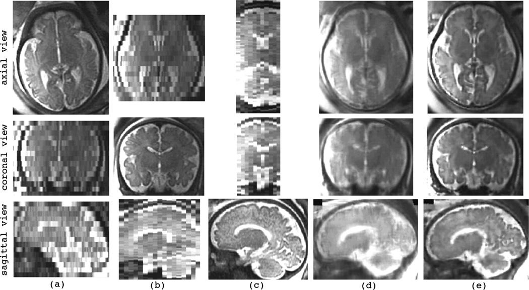
Fast magnetic resonance imaging slice acquisition techniques such as single shot fast spin echo are routinely used in the presence of uncontrollable motion. These techniques are widely used for fetal magnetic resonance imaging (MRI) and MRI of moving subjects and organs. Although high-quality slices are frequently acquired by these techniques, inter-slice motion leads to severe motion artifacts that are apparent in out-of-plane views. Slice sequential acquisitions do not enable 3-D volume representation. In this study, we have developed a novel technique based on a slice acquisition model, which enables the reconstruction of a volumetric image from multiple-scan slice acquisitions. The super-resolution volume reconstruction is formulated as an inverse problem of finding the underlying structure generating the acquired slices. We have developed a robust M-estimation solution which minimizes a robust error norm function between the model-generated slices and the acquired slices. The accuracy and robustness of this novel technique has been quantitatively assessed through simulations with digital brain phantom images as well as high-resolution newborn images. We also report here successful application of our new technique for the reconstruction of volumetric fetal brain MRI from clinically acquired data.


Figures

Fig. 1
Fig. 1
Sample fetal MRI acquired in the axial plane: the three slices on the left show the high in-plane resolution and quality of the slice acquisitions, however, motion can be seen by comparing these three adjacent slices; on the right: sagittal and coronal views of this scan do not reflect the anatomical details due to the fetal motion and the 4 mm slice thickness needed for sufficient signal-to-noise ratio.
Fig. 2
Fig. 2
Algorithmic overview: block diagram of the developed super-resolution reconstruction algorithm; TC stands for the termination condition. Note that the previously developed SDI approaches do not involve the super-resolution reconstruction block. This block aims at minimizing an error norm function between the acquired slices and the estimated volume. This is performed through iterative solutions of Equation (10) and Equation (13).
Fig. 3
Fig. 3
Sample synthetic motion-corrupted slice acquisitions generated from a high-resolution T2-weighted TSE image of a newborn subject. Three views (axial, coronal, sagittal views) of each synthetic image are shown in a column. The slice select direction in (a) and (d) is axial, in (b) and (e) is coronal, and in (c) and (f) is sagittal. No noise or outlier was added to the images in (a) to (c), while images (d) to (f) were contaminated with additive Laplacian noise (power of noise is σ = 200). Image (f) was also corrupted by synthetic slice outliers. Note that the synthetic images involve high-quality slice plane views but exhibit discontinuous tissue boundaries in the out-of-plane views. For example, the synthetic image in (a) with axial slice select direction exhibits sharp anatomic details in the axial view but not in the coronal and sagittal views.
Fig. 4
Fig. 4
The plots of the estimated slice motion parameters (estimated) compared to the actual synthesized slice motion parameters (reference) for the iterative registration and reconstruction of newborn case 2. The six parameters of the randomly generated motion parameters involve three rotations (rx, ry, rz) in degrees and three translations (tx, ty, tz) in millimeters. Each point corresponds to a slice. The points marked by asterisks (*) correspond to the slices detected as moderate and extreme outliers by the box-plot outlier detection method of quartiles based on the MSD of intensity values. Note that the statistical outlier detection method is not perfect and thus a few number of mis-registered slices may not be detected as outliers by MSD, but this analysis indicates that MSD can be used as a relatively reliable measure for assessing the fidelity of slice-to-volume registration.
Fig. 5
Fig. 5
Error image volumes at the beginning and the end of the super-resolution reconstruction algorithm show the convergence of the algorithm; (a) and (b) show the reconstructed error volumes between the estimated volume and the synthesized slice acquisitions. (c) and (d) show the actual error volumes between the estimated volume and the original high-resolution volumes that are available for the validation datasets. Obviously, the algorithm minimizes the error between the estimated volume and the available slice acquisitions (b), but this may not result in perfect match between the reconstructed volume and the original ground truth volume, thus the actual error volume shown in (d) is not as good as (b).
Fig. 6
Fig. 6
The reconstructed volumes for newborn case 2; (a) shows the initial reconstructed volume based on averaging the resampled input scans (AVE), (b) shows the reconstructed volume after ten iterations of slice-to-volume registration and MLE super-resolution reconstruction algorithm, (c) shows the reconstructed volume obtained from one iteration of MLE with known slice motion parameters, (d) shows the original high-resolution TSE volume (used as the ground truth reference for this validation dataset), and (e) shows the reconstructed volume obtained from BSP-SDI with known slice motion parameters. The comparison between BSP-SDI and MLE is considered in section IV-D.
Fig. 7
Fig. 7
The reconstructed volumes for an experiment with mixed noise and slice outliers with the newborn validation dataset; (a) shows the volume reconstructed by ME with l2-norm error function (i.e. ordinary MLE), (b) shows the volume reconstructed by ME with l1-norm error function, and (c) shows the volume reconstructed using the RME technique with Huber’s slice error vector norm function. Severe artifacts appear in the non-robust l2-norm ME volume reconstruction in (a); the black arrows point at some of these artifacts. The volume reconstructed with l1-norm ME in (b) is blurred and still has some artifacts; finally the volume reconstructed with data-adaptive robust RME in (c) is sharp and the effect of slice outliers is appropriately eliminated.
Fig. 8
Fig. 8
Acquired HASTE scans for the volunteer subject experiment; (a)–(c): axial HASTE acquisitions, (d) and (e): coronal HASTE acquisitions, and (f) a sagittal HASTE acquisition. Note that despite the large scale of volunteer motion, the slice quality and resolution is generally preserved in HASTE imaging. However, if the motion is fast, a number of slices may be affected by signal loss and intensity distortion artifacts. A few of these affected slices can be seen in the coronal and sagittal views of the axial acquisition in (b).
Fig. 9
Fig. 9
The reconstructed volumes for the volunteer subject experiment with six HASTE input scans shown in Fig. 8; (a) is the reconstructed volume using LNG-SDI, (b) is the reconstructed volume using the RME technique, (c) is the reference high-resolution TSE volume, and (d) is a TSE volume acquired when the subject was moving in the scanner.
Fig. 10
Fig. 10
The reconstruction of volumetric fetal brain MRI for C6 (GA 19w2d, N = 5); (a)–(c): the acquired SSFSE scans in (a) axial, (b) coronal, and (c) sagittal orthogonal planes, (d) volumetric image reconstructed by LNG-SDI approach, and (e) volumetric image reconstructed by the developed ME technique (with ordinary MLE formulation).
Fig. 11
Fig. 11
The reconstruction of volumetric fetal brain MRI for C12 (GA 36w2d, N = 4); (a)–(c): the acquired SSFSE scans in (a) axial, (b) coronal, and (c) sagittal orthogonal planes, (d) the initial reconstructed volume by averaging (AVE), and (e) the volume reconstructed by the developed RME technique.

References

    1. Prayer D, Brugger P, Prayer L. Fetal MRI: techniques and protocols. Pediatric Radiology. 2004 Sep;vol. 34(no. 9):685–693. - PubMed
    1. Huppert BJ, Brandt KR, Ramin KD, King BF. Single-shot fast spin-echo MR imaging of the fetus: A pictorial essay. Radiographics. 1999;vol. 19(no. suppl 1):S215–S227. - PubMed
    1. Rutherford MA. Magnetic resonance imaging of the fetal brain. Current Opinion in Obstetrics and Gynecology. 2009;vol. 21(no. 2):180–186. - PubMed
    1. Glenn O, Barkovich A. Magnetic resonance imaging of the fetal brain and spine: An increasingly important tool in prenatal diagnosis, part 1. AJNR Am J Neuroradiol. 2006;vol. 27(no. 8):1604–1611. - PMC - PubMed
    1. Vignaux OB, Augui J, Coste J, Argaud C, Le Roux P, Carlier PG, Duboc D, Legmann P. Comparison of single-shot fast spin-echo and conventional spin-echo sequences for MR imaging of the heart: initial experience. Radiology. 2001;vol. 219(no. 2):545–550. - PubMed

Publication types

MeSH terms