. 2010 Aug 6;285(32):24977-86.
doi: 10.1074/jbc.M110.130054. Epub 2010 Jun 7.

Pannexin 2 is expressed by postnatal hippocampal neural progenitors and modulates neuronal commitment

Affiliations

Pannexin 2 is expressed by postnatal hippocampal neural progenitors and modulates neuronal commitment

Leigh Anne Swayne et al. J Biol Chem. .

Abstract

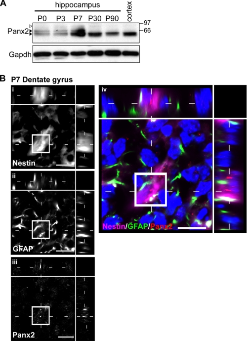
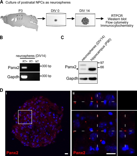
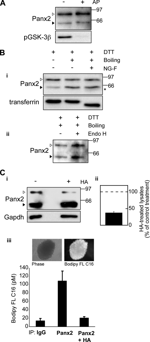
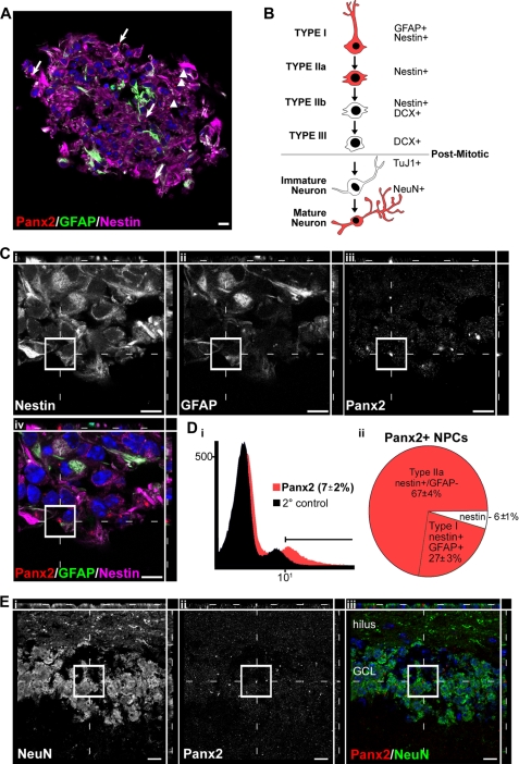
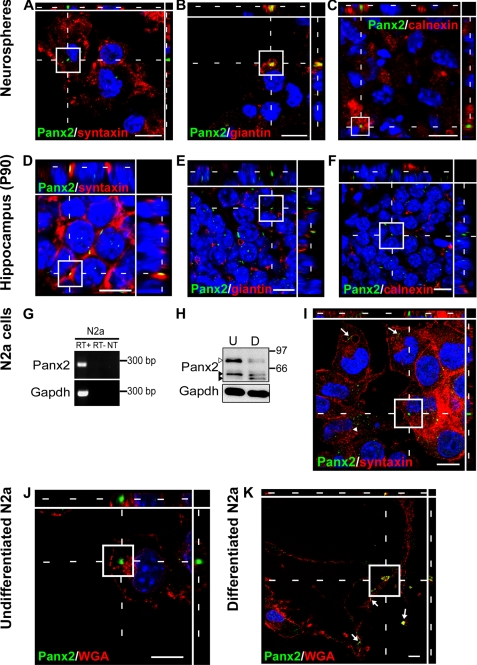
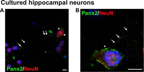
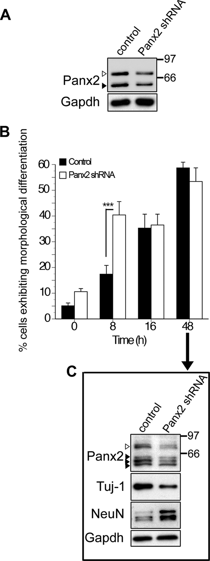
The pannexins (Panx1, -2, and -3) are a mammalian family of putative single membrane channels discovered through homology to invertebrate gap junction-forming proteins, the innexins. Because connexin gap junction proteins are known regulators of neural stem and progenitor cell proliferation, migration, and specification, we asked whether pannexins, specifically Panx2, play a similar role in the postnatal hippocampus. We show that Panx2 protein is differentially expressed by multipotential progenitor cells and mature neurons. Both in vivo and in vitro, Type I and IIa stem-like neural progenitor cells express an S-palmitoylated Panx2 species localizing to Golgi and endoplasmic reticulum membranes. Protein expression is down-regulated during neurogenesis in neuronally committed Type IIb and III progenitor cells and immature neurons. Panx2 is re-expressed by neurons following maturation. Protein expressed by mature neurons is not palmitoylated and localizes to the plasma membrane. To assess the impact of Panx2 on neuronal differentiation, we used short hairpin RNA to suppress Panx2 expression in Neuro2a cells. Knockdown significantly accelerated the rate of neuronal differentiation. Neuritic extension and the expression of antigenic markers of mature neurons occurred earlier in stable lines expressing Panx2 short hairpin RNA than in controls. Together, these findings describe an endogenous post-translational regulation of Panx2, specific to early neural progenitor cells, and demonstrate that this expression plays a role in modulating the timing of their commitment to a neuronal lineage.


Figures

FIGURE 1.
FIGURE 1.
Different Panx2 species are expressed over the course of postnatal hippocampal development. A, three Panx2 species ranging from ∼60 to 85 kDa (arrowheads) are predominant in hippocampal tissue between P0 and P7; two species are detected at P30 (white arrowhead and lower black arrowhead), and a single 60-kDa species predominates at P90, detected also in adult cortex (lower black arrowhead). Membranes were stripped and reprobed for GAPDH as a loading control (lower panel). A representative immunoblot of triplicate experiments is shown. B, Panx2 localizes to neurons (data not shown) and Type I (stem-like) GFAP+/nestin+ NPCs by confocal immunofluorescence of P7 hippocampus. The developing granule cell layer of the dentate gyrus is depicted. Confocal grayscale images with orthogonal views of a triple-labeled Type I NPC are shown in i–iii for each of the primary antibodies used; the pseudocolored merged image is shown in panel iv. Hoechst 33258 was used as a nuclear counterstain (blue). Scale bar, 10 μm. Polyclonal anti-Panx2 (Aviva Systems Biology) was used.
FIGURE 2.
FIGURE 2.
Panx2 is expressed by primary postnatal NPCs cultured as neurospheres. A, schematic of methodology. B, NPCs express Panx2 mRNA. Transcript was detected by reverse transcription-PCR using random-primed RNA extracted from >150 pooled neurosphere cultures. Controls included reactions processed in the absence of reverse transcriptase (RT−) or without template (NT). The same random-primed templates were amplified for GAPDH to confirm template integrity (lower panel). C, Two Panx2 protein species (∼60 kDa (black arrowhead) and ∼85 kDa (white arrowhead)) are detected in neurosphere cultures. The ∼60-kDa species predominates in adult hippocampus. Membranes were stripped and reprobed for GAPDH protein as a loading control (lower panel). A representative immunoblot of triplicate experiments is shown. D, Panx2 expressed in NPC cultures in vitro (i) exhibits a morphology similar to that seen in Type I stem-like NPCs in vivo. The white box indicates the 3× optical zoom inset in ii with orthogonal side views created from a serial confocal z-stack revealing the rod-like Panx2+ structure in greater detail. Three-dimensional reconstructions are provided in supplemental Movies M1 and M2. Hoechst 33258 was used as a nuclear counterstain (blue). Scale bars, 10 μm. Polyclonal anti-Panx2 (Aviva Systems Biology) was used.
FIGURE 3.
FIGURE 3.
NPCs express an S-palmitoylated Panx2 species. Protein lysates were prepared from P3 neurosphere cultures on DIV 14. A, the mobilities of the ∼85-kDa (white arrowhead) and ∼60-kDa species (black arrowhead) were not markedly altered by treatment with alkaline phosphatase (AP). Blots were stripped and reprobed with anti-phospho-GSK3β (Ser9) (pGSK-3β) polyclonal antibody to confirm efficient dephosphorylation (bottom panel). B, the ∼85-kDa species is not N-glycosylated. i, for deglycosylation experiments, lysates were either incubated at 4 °C (first lane) or boiled in the presence of SDS and dithiothreitol (DTT) and incubated at 37 °C with control buffer (second lane) or N-glycosidase F (third lane). Conditions that promote partial depalmitoylation (second and third lanes) induced a shift in the mobility of the ∼85-kDa Panx2 species (white arrowhead) to the ∼60-kDa species (black arrowhead, compare first and second lanes). A small shift to below ∼60 kDa was detected after treatment with N-glycosidase F (asterisk). Blots were stripped and reprobed with anti-transferrin antibody to verify effective deglycosylation (bottom panel). ii, Endo H treatment did not alter Panx2 migration. C, the NPC-specific ∼85-kDa species is a palmitoylated form of Panx2. i, the intensity of the 85-kDa species was markedly reduced following hydroxylamine (HA) treatment. ii, quantitation of this reduction in three independent experiments. Data represent densitometric analysis of the upper band (white arrowhead) standardized to GAPDH and expressed as a percentage of the buffer-treated control. Polyclonal anti-Panx2 (Aviva Systems Biology) was used. iii, to provide direct evidence of palmitoylation, NPCs were metabolically labeled with BODIPY FL hexadecanoic acid (upper panel). Panx2 was immunoprecipitated using the Panx2 C-term polyclonal antibody (Zymed Laboratories Inc./Invitrogen) and treated with buffer or hydroxylamine. Fluorescent palmitate-associated samples after these incubations were extracted and quantified. Data represent pm BODIPY FL hexadecanoic acid extracted from each sample. Panx2 is clearly palmitoylated, and this modification is reversed by treatment with hydroxylamine. All details are as described under “Experimental Procedures.”
FIGURE 4.
FIGURE 4.
Panx2 expression is restricted to Type I and IIa early stem-like hippocampal neural progenitors and mature neurons and is not expressed in intermediate progenitor populations or immature neurons. A, Panx2 localizes to both Type I NPCs (GFAP+/nestin+; arrows) and Type IIa NPCs (GFAP-negative/nestin+; arrowheads) in neurosphere cultures as assessed by confocal microscopy. B, schematic depicting the lineage progression of hippocampal NPCs over the course of postnatal specification to neurons. The antigenic lineage markers used to identify different cell populations by flow cytometry and immunocytochemistry are indicated. C, higher magnification of a Type I cell expressing nestin (i), GFAP (ii), and a Panx2+ rod-like structure (iii). The merged, pseudocolored image is depicted in iv. A Panx2+ Type IIa cell can be seen in the same field (arrowhead). Panx2 was not detected in DCX+ Type IIb or III NPCs or Tuj1+ immature neurons (supplemental Fig. S2). D, the percentage of cells at DIV 14 expressing Panx2 was quantified (i) and characterized (ii) by flow cytometry analysis. E, because the immature neurons generated in our neurosphere cultures do not mature to NeuN+ cells by DIV 14, expression in adult granule neurons was confirmed in P90 hippocampal tissue. Mature NeuN+ hippocampal granule neurons of the dentate gyrus (i) express Panx2 rod-like structures (ii). The pseudocolored merged image is shown in iii. In all micrographs, Hoechst 33258 was used as a nuclear counterstain (blue). Scale bars, 10 μm. Polyclonal anti-Panx2 (Aviva Systems Biology) was used. GCL, granule cell layer.
FIGURE 5.
FIGURE 5.
Panx2 localizes to different subcellular compartments in Type I and IIa NPCs and mature neurons. Subcellular localization was assessed by confocal microcopy using antigenic markers of or carbohydrate-binding proteins labeling plasma membrane (syntaxin, wheat germ agglutinin), Golgi apparatus (giantin), and endoplasmic reticulum (calnexin). A–C, in NPCs, Panx2 does not localize to the plasma membrane (A) but is found at the Golgi apparatus (B) and associated with ER membranes (C). D–F, in mature granule neurons, Panx2 is found at the plasma membrane (D) but not at Golgi (E) or ER (F) membranes. G–K, this shift in localization is associated with neuronal differentiation. Panx2 mRNA (G, reverse transcription-PCR) and protein (H, Western analysis; I–K, confocal immunofluorescence) are expressed by undifferentiated and differentiated N2a cells (U, undifferentiated; D, differentiated). Both the 85-kDa palmitoylated (white arrowhead) and 60-kDa unpalmitoylated (upper black arrowhead) species are present in undifferentiated N2a cells with intensity of the 85-kDa form reduced following differentiation. A novel Panx2 species is also detected (lower black arrowhead) (H). Abbreviations and controls are as described in the legend to Fig. 2. J and K, further subcellular localization comparisons were made using fluorescently labeled wheat germ agglutinin as a plasma membrane marker. In undifferentiated N2a cells, Panx2 is not detected at the plasma membrane (J). Following 48 h of differentiation with 10 μm retinoic acid, Panx2 colocalizes with wheat germ agglutinin at the plasma membrane in neuritic extensions (K, arrows). This shift in localization is associated with a reduction in Panx2 protein expression, in particular the ∼85-kDa species (white arrowhead, H). In all micrographs, Hoechst 33258 was used as a nuclear counterstain (blue). Scale bars, 10 μm. Polyclonal anti-Panx2 (Aviva Systems Biology) was used.
FIGURE 6.
FIGURE 6.
Panx2 localizes to the plasma membrane of primary hippocampal neurons. A and B, confocal images of hippocampal neuronal cultures immunolabeled for the primarily nuclear marker NeuN and Panx2. The arrows indicate Panx2 present at the plasma membrane of cell soma and along process varicosities. Hoechst 33258 was used as a nuclear counterstain (blue). Scale bars, 10 μm. Polyclonal anti-Panx2 (Aviva Systems Biology) was used.
FIGURE 7.
FIGURE 7.
shRNA-mediated knockdown of Panx2 accelerates morphological and biochemical indices of differentiation. A, Western blot of lysates from N2a cells stably transfected with empty vector (first lane) or plasmid encoding Panx2 shRNA (second lane). A knockdown of about 50% is observed in both the ∼85-kDa (white arrowhead) and ∼60-kDa (black arrowhead) Panx2 species. B and C, empty vector controls and Panx2 shRNA-expressing N2a cells were differentiated for up to 48 h with retinoic acid (10 μm) as described under “Experimental Procedures.” Cells were defined as morphologically differentiated if they exhibited one or more neurites of a length greater than the diameter of the cell soma. B, morphological indices of neuronal differentiation appeared earlier when Panx2 expression was suppressed (p < 0.001, two-way analysis of variance, post hoc Bonferroni). C, after 48 h of differentiation, N2a cells expressed multiple Panx2 species reduced in shRNA expressing cells (arrowheads). The white arrowhead indicates the ∼85-kDa species. Although 60% of cells were morphologically differentiated in both groups by this time point (C), N2a cells expressing Panx2 shRNA exhibited higher levels of the mature neuronal antigen, NeuN, and lower levels of Tuj1-reactive βIII tubulin expressed by immature neurons than control cells, indicative of a greater degree of biochemical maturation. Polyclonal anti-Panx2 (Aviva Systems Biology) was used. Error bars, S.E.

References

    1. Kempermann G., Jessberger S., Steiner B., Kronenberg G. (2004) Trends Neurosci. 27, 447–452 - PubMed
    1. Li G., Pleasure S. J. (2010) Curr. Opin. Neurobiol. 20, 126–133 - PMC - PubMed
    1. Ma D. K., Bonaguidi M. A., Ming G. L., Song H. (2009) Cell Res. 19, 672–682 - PMC - PubMed
    1. Thompson A., Boekhoorn K., Van Dam A. M., Lucassen P. J. (2008) Genes Brain Behav. 7, Suppl. 1, 28–42 - PubMed
    1. D'Ascenzo M., Piacentini R., Casalbore P., Budoni M., Pallini R., Azzena G. B., Grassi C. (2006) Eur. J. Neurosci. 23, 935–944 - PubMed

Publication types