

A soft kinetic data structure for lesion border detection
- PMID: 20529909
- PMCID: PMC2881363
- DOI: 10.1093/bioinformatics/btq178
A soft kinetic data structure for lesion border detection
Abstract
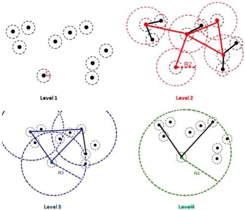
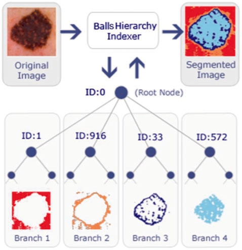
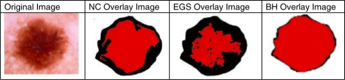
Motivation: The medical imaging and image processing techniques, ranging from microscopic to macroscopic, has become one of the main components of diagnostic procedures to assist dermatologists in their medical decision-making processes. Computer-aided segmentation and border detection on dermoscopic images is one of the core components of diagnostic procedures and therapeutic interventions for skin cancer. Automated assessment tools for dermoscopic images have become an important research field mainly because of inter- and intra-observer variations in human interpretations. In this study, a novel approach-graph spanner-for automatic border detection in dermoscopic images is proposed. In this approach, a proximity graph representation of dermoscopic images in order to detect regions and borders in skin lesion is presented.
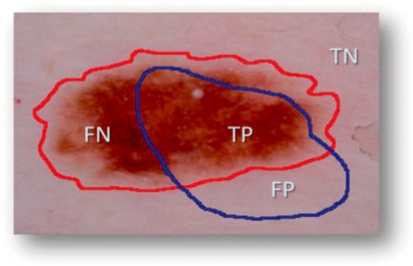
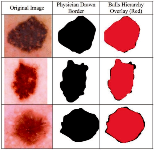
Results: Graph spanner approach is examined on a set of 100 dermoscopic images whose manually drawn borders by a dermatologist are used as the ground truth. Error rates, false positives and false negatives along with true positives and true negatives are quantified by digitally comparing results with manually determined borders from a dermatologist. The results show that the highest precision and recall rates obtained to determine lesion boundaries are 100%. However, accuracy of assessment averages out at 97.72% and borders errors' mean is 2.28% for whole dataset.
Figures
References
-
- Alexandron G, et al. Kinetic and dynamic data structures for convex hulls and upper envelopes. Comput. Geometr. Theory Appl. 2007;36:144–158.
-
- Argenziano G, et al. Dermoscopy: a Tutorial. Milan, Italy: EDRA Medical Publishing and New Media; 2002.
-
- Basch J, et al. Data structures for mobile data. J. Algorithms. 1999;31:1–28.
-
- Binder M, et al. Epiluminescence microscopy. A useful tool for the diagnosis of pigmented skin lesions for formally trained dermatologists. Arch. Dermatol. 1995;31:286–291. - PubMed
MeSH terms
LinkOut - more resources
Full Text Sources
Medical
