. 2010 Jun 15;26(12):i57-63.
doi: 10.1093/bioinformatics/btq219.

As-rigid-as-possible mosaicking and serial section registration of large ssTEM datasets

Affiliations

As-rigid-as-possible mosaicking and serial section registration of large ssTEM datasets

Stephan Saalfeld et al. Bioinformatics. .

Abstract

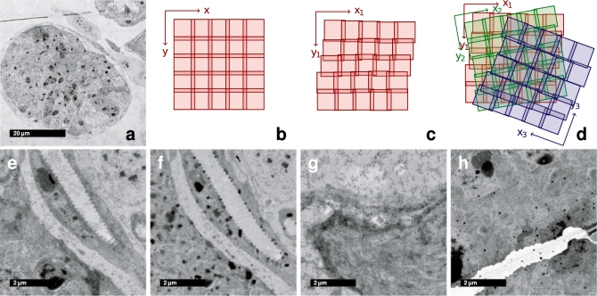
Motivation: Tiled serial section Transmission Electron Microscopy (ssTEM) is increasingly used to describe high-resolution anatomy of large biological specimens. In particular in neurobiology, TEM is indispensable for analysis of synaptic connectivity in the brain. Registration of ssTEM image mosaics has to recover the 3D continuity and geometrical properties of the specimen in presence of various distortions that are applied to the tissue during sectioning, staining and imaging. These include staining artifacts, mechanical deformation, missing sections and the fact that structures may appear dissimilar in consecutive sections.

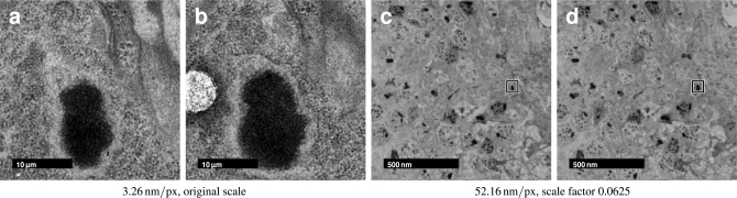
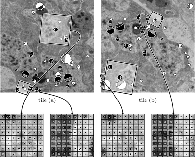
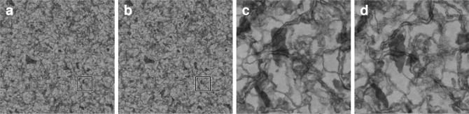
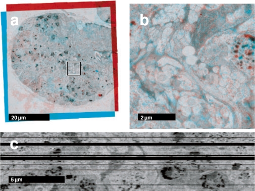
Results: We developed a fully automatic, non-rigid but as-rigid-as-possible registration method for large tiled serial section microscopy stacks. We use the Scale Invariant Feature Transform (SIFT) to identify corresponding landmarks within and across sections and globally optimize the pose of all tiles in terms of least square displacement of these landmark correspondences. We evaluate the precision of the approach using an artificially generated dataset designed to mimic the properties of TEM data. We demonstrate the performance of our method by registering an ssTEM dataset of the first instar larval brain of Drosophila melanogaster consisting of 6885 images.

Availability: This method is implemented as part of the open source software TrakEM2 (http://www.ini.uzh.ch/~acardona/trakem2.html) and distributed through the Fiji project (http://pacific.mpi-cbg.de).


Figures

Fig. 1.
Fig. 1.
Properties of ssTEM image mosaics. (a) Low magnification overview of a single section mosaic consisting of 9 × 9 registered tiles. (b) Each section is imaged as a sequence of overlapping tiles that is assumed to be a regular grid. (c) Each tile's true pose in the local section coordinate frame is affected by odometry errors of the microscope, that are propagated over consecutive tiles. (d) The relative pose of two consecutive sections is arbitrarily shifted and rotated. Clean sections are relatively rare (e); typically sections contain artifacts such as dirt (f), staining precipitate (g) and folds or cracks (h).
Fig. 2.
Fig. 2.
Similarity of serial sections depends on the scale of observation. Two consecutive 60 nm sections from the registered Drosophila first instar larval brain dataset showing a part of the cortex and the neuropile. The same region is shown with a resolution of 3.26 nm/px (a,b) and 52.16 nm/px (c,d). It is clearly visible, that the latter, having approximately isotropic resolution, shows significant similarity across sections while it is hard to identify corresponding pixels in the higher resolution example.
Fig. 3.
Fig. 3.
SIFT-feature correspondences in two overlapping tiles from adjacent sections. 1003 feature candidates were extracted in tile (a), 971 in tile (b). 41 correspondence pairs were identified by local feature descriptor matching, 19 of them are true matches consistent to common transformation model. False matches are displayed in white, true matches in black. The size of the circles is proportional to the features scale, the filled part visualizes a feature's orientation. Two true correspondence pairs are selected as an example for the local SIFT-descriptor. The values of the local histogram bins are shown as combs on top of the local region the descriptor is extracted from.
Fig. 4.
Fig. 4.
Artificially generated evaluation dataset. The dataset simulates thin transilluminated volumes of 20 px thickness with membrane- and blob-like structures at various scales. Structures are defined by volumetric density functions. Locations with higher density scatter more ‘light’ than those with lower density and appear darker in the shadow projection. Two adjacent sections are shown to visualize the cross-section change of visible structures. In (a,b), the full dataset (4096 × 4096 px) is shown at low magnification, an area of 512 × 512 px is marked and shown in (c,d) at high magnification.
Fig. 5.
Fig. 5.
Drosophila first instar larval brain ssTEM dataset. Two consecutive registered sections from the dataset as red-cyan color merge. The diameter of the brain is ∼60 μm. (a) The section mosaics as a whole with the areas zoomed in (b) marked. (c) A single perpendicular section through the entire registered volume. Several sections were lost during sectioning and collecting onto the electron microscopy grid and shown here as black rows.

Similar articles

Cited by

References

    1. Anderson JR, et al. A computational framework for ultrastructural mapping of neural circuitry. PLoS Biol. 2009;7:e1000074. - PMC - PubMed
    1. Bay H, et al. SURF: Speeded up robust features. Comput. Vis. Image Understand. 2008;110:346–359.
    1. Brown M, Lowe DG. ICCV '03: Proceedings of the Ninth IEEE International Conference on Computer Vision. Washington, DC: IEEE Computer Society; 2003. Recognising panoramas; pp. 1218–1225.
    1. Cardona A. Proceedings of the 1st ImageJ User and Developer Conference. Luxembourg: 2006. TrakEM2: an ImageJ-based program for morphological data mining and 3D modeling; pp. 18–19. May.
    1. Cardona A, et al. Drosophila brain development: Closing the gap between a macroarchitectural and microarchitectural approach. Cold Spring Harb. Symp. Quant. Biol. 2009 [Epub ahead of print, doi:10.1101/sqb.2009.74.037] - PMC - PubMed

Publication types

MeSH terms