

Functional radiographic diagnosis of the lumbar spine. Flexion-extension and lateral bending
- PMID: 2052999
- PMCID: PMC8009657
- DOI: 10.1097/00007632-199105000-00014
Functional radiographic diagnosis of the lumbar spine. Flexion-extension and lateral bending
Abstract
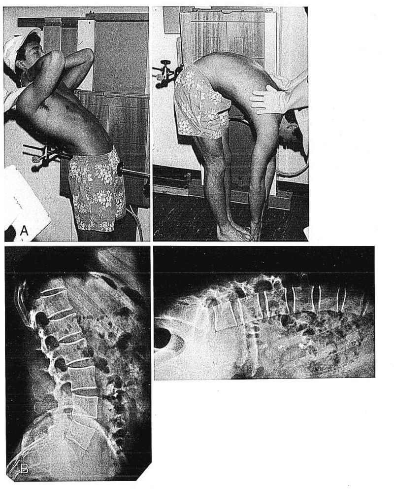
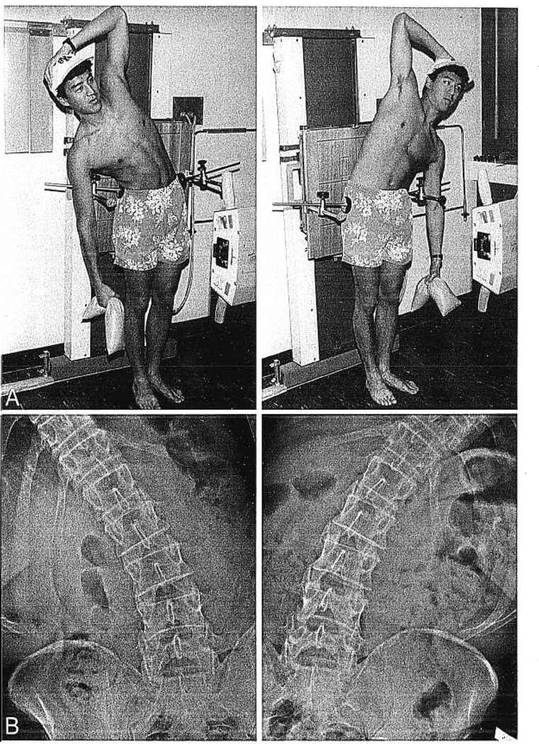
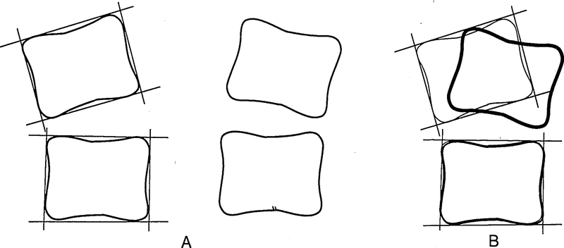
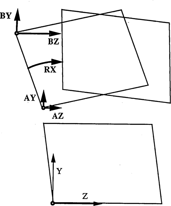
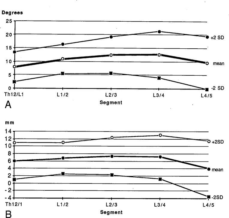
Several attempts have been made to measure the segmental range of motion in the lumbar spine during flexion-extension with the purpose of gathering additional data for the diagnosis of instability. The previous studies were performed in vitro or in vivo during active motion. The aim of this study was to obtain normal values of passively performed segmental motions. Forty-one healthy adults were examined by means of functional radiographs during flexion-extension and lateral bending. A graphic construction method and a computer-assisted method were used to measure rotations. Comparing with recent in vivo studies, the values obtained for normal angles of rotation were predominately larger. This might be due to the passive examination used in the study. The graphic construction method and computer-assisted method techniques are equally reliable, but the computer-assisted method method yields other important kinematic data, such as translations. It is proposed that passive motion be applied during functional examination of patients with suspected instabilities. However, the large variation of rotational values between individuals in the normal population may limit the clinical usefulness of functional lumbar analysis using this parameter. Future studies should explore the clinical relevance of determining altered segmental mobility in low-back pain patients.
Figures
References
-
- Allbrook D: Movements of the lumbar spinal column. J Bone Joint Surg 39B:339–345, 1957 - PubMed
-
- Begg AC, Falconer MA: Plain radiography in intraspinal protrusion of lumbar intervertebral discs: A correlation with operative findings. Br J Surg 36:225–239, 1949 - PubMed
-
- Clayson SJ, Newman IM, Debevec DF, et al. : Evaluation of mobility of hip and lumbar vertebrae of normal young women. Arch Phys Med 43:1–8, 1962 - PubMed
-
- Dvorak J, Fröhlich D, Penning L, Baumgaertner H, Panjabi MM: Functional radiographic diagnosis of the cervical spine: fLexion/extension. Spine 13:748–755, 1988 - PubMed
-
- Dimnet J, Fischer LP, Gonon G, Carret JP: Radiographic studies of lateral flexion in the lumbar spine. J Biomech 11:143–150, 1978 - PubMed
Publication types
MeSH terms
Grants and funding
LinkOut - more resources
Full Text Sources