Comparative Study
. 2010 Aug 1;588(Pt 15):2891-903.
doi: 10.1113/jphysiol.2010.190504. Epub 2010 Jun 7.

Length dependence of force generation exhibit similarities between rat cardiac myocytes and skeletal muscle fibres

Affiliations
Comparative Study

Length dependence of force generation exhibit similarities between rat cardiac myocytes and skeletal muscle fibres

Laurin M Hanft et al. J Physiol. .

Abstract

According to the Frank-Starling relationship, increased ventricular volume increases cardiac output, which helps match cardiac output to peripheral circulatory demand. The cellular basis for this relationship is in large part the myofilament length-tension relationship. Length-tension relationships in maximally calcium activated preparations are relatively shallow and similar between cardiac myocytes and skeletal muscle fibres. During twitch activations length-tension relationships become steeper in both cardiac and skeletal muscle; however, it remains unclear whether length dependence of tension differs between striated muscle cell types during submaximal activations. The purpose of this study was to compare sarcomere length-tension relationships and the sarcomere length dependence of force development between rat skinned left ventricular cardiac myocytes and fast-twitch and slow-twitch skeletal muscle fibres. Muscle cell preparations were calcium activated to yield 50% maximal force, after which isometric force and rate constants (k(tr)) of force development were measured over a range of sarcomere lengths. Myofilament length-tension relationships were considerably steeper in fast-twitch fibres compared to slow-twitch fibres. Interestingly, cardiac myocyte preparations exhibited two populations of length-tension relationships, one steeper than fast-twitch fibres and the other similar to slow-twitch fibres. Moreover, myocytes with shallow length-tension relationships were converted to steeper length-tension relationships by protein kinase A (PKA)-induced myofilament phosphorylation. Sarcomere length-k(tr) relationships were distinct between all three cell types and exhibited patterns markedly different from Ca(2+) activation-dependent k(tr) relationships. Overall, these findings indicate cardiac myocytes exhibit varied length-tension relationships and sarcomere length appears a dominant modulator of force development rates. Importantly, cardiac myocyte length-tension relationships appear able to switch between slow-twitch-like and fast-twitch-like by PKA-mediated myofibrillar phosphorylation, which implicates a novel means for controlling Frank-Starling relationships.


Figures

Figure 1
Figure 1. Simultaneous measurement of force at three different sarcomere lengths in a cardiac myocyte preparation
Top right, a photomicrograph of a cardiac myocyte at sarcomere length 2.30 μm and an illustration of the permeabilized muscle cell attachment apparatus. The rest of the figure shows a slow-time base sarcomere length recording using an IonOptix SarcLen system (IonOptix, Milton, MA, USA) and the corresponding (fast-time base) force traces at sarcomere lengths 2.30 μm, 2.15 μm and 1.95 μm during submaximal Ca2+ activation (pCa 5.7) of a rat cardiac myocyte preparation. Upper left force trace is during maximal calcium activation (pCa 4.5) at sarcomere length 2.30 μm.
Figure 2
Figure 2. Sarcomere length–tension relationships for rat fast-twitch skeletal muscle fibres, slow-twitch skeletal muscle fibres and cardiac myocytes
Cardiac myocyte preparations exhibited two populations of length–tension relationships, one steeper even than fast-twitch fibres and the other similar to slow-twitch fibres. Inset shows passive length–tension relationships of rat skinned cardiac myocytes, fast-twitch skeletal muscle fibres, and slow-twitch skeletal muscle fibres. The steepness of active length–tension relationships was independent of passive tension. Slopes of length–tension relationships were: fast-twitch fibres: 1.62 ± 0.17*# (n= 10); slow-twitch fibres: 1.02 ± 0.12§ (n= 8); cardiac myocytes (steep population): 1.85 ± 0.16*#§ (n= 6); cardiac myocytes (shallow population): 1.09 ± 0.15§ (n= 13); *P < 0.05 vs. slow-twitch fibres, #P < 0.05 vs. cardiac myocyte shallow population, §P < 0.05 vs. fast-twitch fibres.
Figure 3
Figure 3. Effects of PKA on cardiac myocyte length–tension relationships
A, an autoradiogram showing radiolabelled phosphate incorporation into rat cardiac myofibrillar proteins (MyBP-C and cTnI) upon PKA treatment. Lanes 1 and 2 contain permeabilized cardiac myocytes in the presence of [32P]ATP either without (lane 1) or with PKA (lane 2). B, cardiac myocyte force traces during submaximal Ca2+ activations at four different sarcomere lengths before and after PKA treatment. C, normalized cardiac myocyte sarcomere length–tension relationships before and after PKA treatment (n= 6). PKA-induced phosphorylation markedly steepened the length–tension relationship in these preparations. Left inset shows a representative experiment whereby PKA treatment did not alter the length–tension curve of a cardiac myocyte preparation that had a steep length–tension relationship. Right inset shows a control experiment whereby the addition of PKA in the presence of protein kinase inhibitor (PKI) did not alter the length–tension relationship. The bottom inset autoradiogram shows enhanced PKA-induced phosphate incorporation into cardiac myofilament proteins from rats treated with propranolol (lane 2) versus control rats (lane 1). PKI markedly inhibited the PKA-induced phosphate incorporation into cardiac myofibrillar proteins (lane 3).
Figure 4
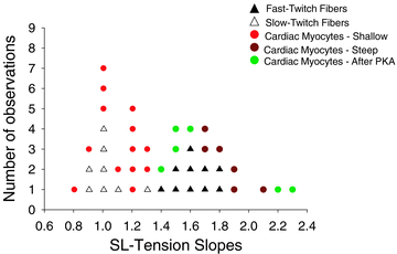
Figure 4. Histogram of slopes of sarcomere length–tension relationships
The histogram indicates two populations of sarcomere length–tension relationships in striated muscle. One population had shallow slopes (ranging from 0.80 to 1.30) and consisted of slow-twitch skeletal muscle fibres and a subgroup of cardiac myocytes. The other population consisted of fast-twitch fibres and a different subgroup of cardiac myocytes that had steeper slopes that ranged from 1.40 to 2.40.
Figure 5
Figure 5. Autoradiogram showing PKA-mediated phosphorylation of MyBP-C and cTnI of myofibrils from different regions of rat ventricular free wall
PKA-induced back-phosphorylation of myofibrils did not show any differences from myocytes isolated from the epicardium, mid-wall, or endocardial regions of the left ventricle.
Figure 6
Figure 6. Sarcomere length dependence of rate constant of force redevelopment (ktr)
A, sarcomere length dependence of ktr for fast-twitch skeletal muscle fibres, slow-twitch fibres and the two populations of cardiac myocyte preparations, one that displayed steep and the other that displayed shallow length–tension relationships. Sarcomere length dependence of ktr was unique for each of the different muscle cell preparations and ktr seemed to increase at sarcomere lengths below ∼1.85 μm in all the preparations. This occurred despite reductions in force, implicating that sarcomere length overrides the activation dependence of ktr normally observed in striated muscle preparations (Gordon et al. 2000). B, sarcomere length dependence of ktr in cardiac myocyte preparations before and after PKA treatment. PKA decreased ktr at all sarcomere lengths and prevented the up-turn in ktr sarcomere lengths below ∼1.85 μm observed in all other preparations. (Data points are means ±s.e.m.)

Similar articles

Cited by

References

    1. Adhikari BB, Regnier M, Rivera AJ, Kreutziger KL, Martyn DA. Cardiac length dependence of force and force redevelopment kinetics with altered cross-bridge cycling. Biophys J. 2004;87:1784–1794. - PMC - PubMed
    1. Ait mou Y, le Guennac J-Y, Mosca E, de Tombe PP, Cazorla O. Differential contribution of cardiac sarcomeric proteins in the myofibrillar force response to stretch. Pflugers Arch. 2008;457:25–36. - PMC - PubMed
    1. Allen JD, Moss RL. Factors influencing the ascending limb of the sarcomere length-tension relationship in rabbit skinned muscle fibers. J Physiol. 1987;390:119–136. - PMC - PubMed
    1. Bardswell SC, Kentish J. Resting and active sarcomere lengths in isolated rat ventricular myocytes. Biophys J. 2003;84:431a.
    1. Braunwald E, Ross J. Applicability of Starling's law of the heart to man. Circ Res. 1964;Suppl ii:169–181. - PubMed

Publication types

LinkOut - more resources