. 2010 Aug;30(16):4077-91.
doi: 10.1128/MCB.01806-08. Epub 2010 Jun 7.

Epidermis-type lipoxygenase 3 regulates adipocyte differentiation and peroxisome proliferator-activated receptor gamma activity

Affiliations

Epidermis-type lipoxygenase 3 regulates adipocyte differentiation and peroxisome proliferator-activated receptor gamma activity

Philip Hallenborg et al. Mol Cell Biol. 2010 Aug.

Abstract

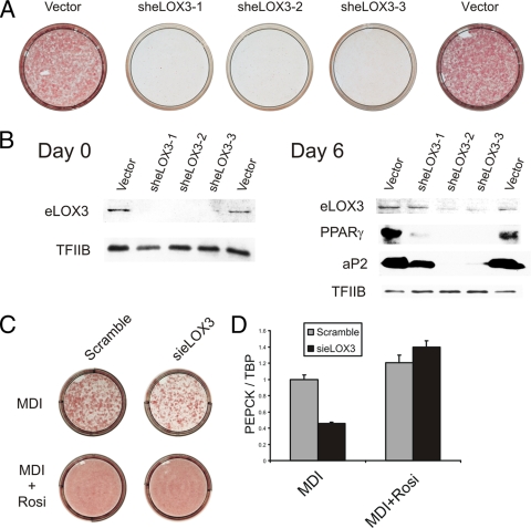
The nuclear receptor peroxisome proliferator-activated receptor gamma (PPAR gamma) is essential for adipogenesis. Although several fatty acids and their derivatives are known to bind and activate PPAR gamma, the nature of the endogenous ligand(s) promoting the early stages of adipocyte differentiation has remained enigmatic. Previously, we showed that lipoxygenase (LOX) activity is involved in activation of PPAR gamma during the early stages of adipocyte differentiation. Of the seven known murine LOXs, only the unconventional LOX epidermis-type lipoxygenase 3 (eLOX3) is expressed in 3T3-L1 preadipocytes. Here, we show that forced expression of eLOX3 or addition of eLOX3 products stimulated adipogenesis under conditions that normally require an exogenous PPAR gamma ligand for differentiation. Hepoxilins, a group of oxidized arachidonic acid derivatives produced by eLOX3, bound to and activated PPAR gamma. Production of hepoxilins was increased transiently during the initial stages of adipogenesis. Furthermore, small interfering RNA-mediated or retroviral short hairpin RNA-mediated knockdown of eLOX3 expression abolished differentiation of 3T3-L1 preadipocytes. Finally, we demonstrate that xanthine oxidoreductase (XOR) and eLOX3 synergistically enhanced PPAR gamma-mediated transactivation. Collectively, our results indicate that hepoxilins produced by the concerted action of XOR and eLOX3 may function as PPAR gamma activators capable of promoting the early PPAR gamma-dependent steps in the conversion of preadipocytes into adipocytes.


Figures

FIG. 1.
FIG. 1.
Forced expression of eLOX3 increases differentiation of Rb/ MEFs. Cells were transduced with either an empty or eLOX3-expressing vector and selected using puromycin. Cells were induced to differentiate according to the MDI protocol with the inclusion of either vehicle, 20 μM baicalein, or 20 μM baicalein and 0.5 μM rosiglitazone from time of induction. Eight days after induction, cells were scored for differentiation. (A) Cells stained for lipid accumulation using oil red O. (B) Expression of adipocyte marker genes, PPARγ2 and aP2, was analyzed by Western blotting. (C) Protein levels of eLOX3 in wild-type and Rb/ MEFs at the onset of differentiation as analyzed by Western blotting. TFIIB was included as a loading control. One representative experiment out of three independent experiments performed in duplicate is shown.
FIG. 2.
FIG. 2.
The enzymatic activity of eLOX3 is required for its adipogenic effect. Rb/ MEFs were transduced with vectors expressing eLOX3, the eLOX3Δ inactive mutant, or vector control and selected using puromycin. Eight days after induction with MDI, cells were scored for adipogenic conversion. (A) Cells were stained for lipid accumulation using oil red O. (B) Expression of adipocyte marker genes, PPARγ and aP2, was analyzed by Western blotting. Data are representative of three independent experiments performed in duplicate. (C) Enzymatic properties of the eLOX3Δ mutant. Cells were transiently transfected with wild-type eLOX3, eLOX3Δ, or LacZ control expression vectors, lysed in LOX assay buffer, and incubated with 100 μM 15(S)-HPETE for 15 min at 37°C. Products were extracted and run on a high-performance liquid chromatograph, and eluates were monitored at 205 nM. Depicted is conversion of 15(S)-HPETE into 13(R)-hydroxy-14(S),15(S)-HepEtrE with the activity of wild-type eLOX3 set to 100%. Data are representative of two independent experiments. (D) Baicalein inhibits enzymatic activity of known LOXs. Expression vectors harboring cDNAs encoding different LOXs were transiently transfected into 293 cells. Cells were lysed in LOX assay buffer and incubated with appropriate substrate and increasing amounts of baicalein. Enzymatic activation is depicted as percent activity with activation in the absence of baicalein set to 100%. (E and F) Identified products of eLOX3, HXA3 and HXB3, increase adipose conversion of Rb/ MEFs. Cells were induced to differentiate according to the MDI protocol with addition of 10 μM HXA3 or HXB3 or vehicle control at the time of induction and on days 1, 2, and 3. Adipogenic conversion was investigated 8 days after induction. (E) Morphological differentiation and staining of lipids by oil red O. (F) Expression of adipogenic marker genes by real-time PCR. PEPCK, phosphoenolpyruvate carboxykinase. One representative experiment out of three independent experiments performed in duplicate is shown.
FIG. 3.
FIG. 3.
eLOX3 and its products induce adipogenesis in 3T3-L1 preadipocytes. Cells were transduced with either empty vector or eLOX3-expressing vector and selected using puromycin. Cells were induced to differentiate with dexamethasone and insulin. Eight days after induction, cells were scored for differentiation as measured by oil red O staining (A). (B and C) 3T3-L1 cells were induced to differentiate with dexamethasone and insulin and with addition of HXA3, HXB3, rosiglitazone, or vehicle control at the time of induction and on days 1, 2, and 3. Adipogenic conversion was investigated 8 days after induction. (B) Morphological differentiation and staining of lipids by oil red O. (C) Expression of adipogenic marker genes by real-time PCR. Data are represented as induction relative to the induction by rosiglitazone over vehicle control. Induction by rosiglitazone is set to 100%. PEPCK, phosphoenolpyruvate carboxykinase. (D) LC/MS analyses of conditioned medium samples from 3T3-L1 cells treated with either DI or MDI for 24 h. Only peaks from signals corresponding to species with a molecular mass similar to that of hepoxilin (335.1 Da) are shown. The peak from the eluted hepoxilin is marked by a red arrow. (E) MS/MS analyses of hepoxilin standards (left and middle panels) and the coeluting peak from day 1 medium from 3T3-L1 cells induced to differentiate with MDI. One representative experiment out of two independent experiments performed in duplicate is shown.
FIG. 4.
FIG. 4.
Hepoxilin A3 and B3 bind and activate PPARγ. (A) HXA3 and HXB3 activate PPARγLBD in a dose-dependent manner. Rb/ MEFs transfected with a Gal4-DBD-PPARγ-LBD expression vector and a UASGal-TK-luciferase reporter were treated with vehicle, 0.5 μM rosiglitazone, or increasing concentrations of HXA3 or HXB3. Luciferase activity was normalized to ß-galactosidase activity and is presented as fold change over vehicle only control. One representative experiment out of four independent experiments performed in triplicate is shown. (B) HXA3, HXB3, and rosiglitazone displace a fluorescent ligand from the PPARγ-LBD. RFU, relative fluorescence units. One representative experiment out of three independent experiments performed in triplicate is shown. (C) Rb−/− MEFs transfected with pDR1 Luc and either PPARγ, PPARγ Q286P, or empty vector treated with 1 μM rosiglitazone, 10 μM HXA3, or 10 μM HXB3. Luciferase activity was normalized to β-galactosidase activity. Data represent three independent experiments performed in triplicate. (D) Potentiation of insulin-stimulated glucose uptake by vehicle, rosiglitazone, HXA3, or HXB3. Differentiated 3T3-L1s were treated with the indicated compounds, and basal or insulin-stimulated uptake of radioactively labeled glucose was measured. Data are presented as fold uptake over the vehicle only control. Data represent two independent experiments performed in octuplicate. (E) GST-PPARγ DE pulldown of TRAP220 in the presence of vehicle, 1 μM rosiglitazone, 10 μM HXA3, or 10 μM HXB3. One representative experiment out of two independent experiments is shown. (F) 3T3-L1 cells were transfected with GFP-PPARγ2 and myc-eLOX3, and localization of the two fusion proteins was determined by confocal microscopy.
FIG. 5.
FIG. 5.
Knockdown of eLOX3 expression prevents adipocyte differentiation of 3T3-L1 cells. Cells were transduced with shRNA constructs against eLOX3, selected with puromycin, and induced to differentiate according to the MDI protocol. (A) Cells stained with oil red O. (B) Detection of eLOX3 expression and adipocyte markers using Western blotting. TFIIB was included as a loading control. One representative experiment out of three independent experiments performed in duplicate is shown. (C and D) 3T3-L1 preadipocytes were transfected with either scrambled or eLOX3 RNAi using DeliverX Plus and induced to differentiate in the presence or absence of 0.5 μM rosiglitazone. (C) Cells were stained for lipid accumulation using oil red O. (D) Expression of PEPCK mRNA as a marker for differentiation was examined by real-time PCR. One representative experiment out of two independent experiments performed in triplicate is shown.
FIG. 6.
FIG. 6.
eLOX3 cooperates with XOR in the activation of PPARγ. (A) 15(S)- and 12(S)-HETE in the medium from wild-type MEFs before (day 0) and 24 h after (day 1) induction with MDI. Data represent two independent experiments performed in duplicate. (B) Rb/ MEFs were transiently transfected using UASGal-TK-luciferase reporter, Gal4-DBD-PPARγ-LBD, and either XOR, eLOX3, or both. Transfected cells were stimulated with 5 μM arachidonic acid. Luciferase activity was normalized to ß-galactosidase activity and is presented as fold change over vector only control. One representative experiment out of four independent experiments performed in triplicates is shown. (C) Expression of XOR in wild-type and Rb/ MEFs was analyzed by real-time PCR and normalized to TBP expression. Data represent two independent experiments performed in triplicate.
FIG. 7.
FIG. 7.
The antioxidant NAC abolishes adipose conversion. (A and B) 3T3-L1 preadipocytes were induced to differentiate according to the MDI protocol with the inclusion of rosiglitazone (0.5 μM) and/or NAC (5 mM) or vehicle alone. Differentiation was determined by oil red O staining (A) or expression of adipocyte marker genes (B). ACC, acetyl coenzyme A carboxylase; HSL, hormone-sensitive lipase; GLUT4, facilitated glucose transporter, member 4. Data represent two independent experiments performed in duplicate. (C and D) 3T3-L1 preadipocytes were induced to differentiate according to the MDI protocol with the inclusion of rosiglitazone (0.5 μM) and/or NAC (5 mM) or vehicle alone from day 0 to day 4. Differentiation was determined by oil red O staining (C) and expression of adipocyte marker genes (D). Data represent two independent experiments performed in duplicate. (E) 3T3-L1 cells were induced to differentiate in the presence and absence of 5 mM NAC. Expression of C/EBPβ and XOR was analyzed at indicated time points. Data represent two independent experiments performed in duplicate.
FIG. 8.
FIG. 8.
Hypothetical pathway for the generation of PPARγ ligands during the early stages of adipocyte differentiation. AA, arachidonic acid; MDA, malondialdehyde; 4-HNE, 4-hydroxy-nonenals.

Similar articles

Cited by

References

    1. Araujo, P., Z.-Y. Du, T.-T. Nguyen, and E. Holen. 2008. Direct injection of redissolved cell culture media into a single-column liquid chromatography coupled to mass spectrometry for the measurement of PGE2. Open Anal. Chem. J. 2:62-66.
    1. Baker, P. R., F. J. Schopfer, S. Sweeney, and B. A. Freeman. 2004. Red cell membrane and plasma linoleic acid nitration products: synthesis, clinical identification, and quantitation. Proc. Natl. Acad. Sci. U. S. A. 101:11577-11582. - PMC - PubMed
    1. Barak, Y., M. C. Nelson, E. S. Ong, Y. Z. Jones, P. Ruiz-Lozano, K. R. Chien, A. Koder, and R. M. Evans. 1999. PPAR gamma is required for placental, cardiac, and adipose tissue development. Mol. Cell 4:585-595. - PubMed
    1. Bastie, C., D. Holst, D. Gaillard, C. Jehl-Pietri, and P. A. Grimaldi. 1999. Expression of peroxisome proliferator-activated receptor PPARdelta promotes induction of PPARγ and adipocyte differentiation in 3T3C2 fibroblasts. J. Biol. Chem. 274:21920-21925. - PubMed
    1. Bastie, C., S. Luquet, D. Holst, C. Jehl-Pietri, and P. A. Grimaldi. 2000. Alterations of peroxisome proliferator-activated receptor delta activity affect fatty acid-controlled adipose differentiation. J. Biol. Chem. 275:38768-38773. - PubMed

Publication types

MeSH terms