. 2010 Aug;31(8):1360-6.
doi: 10.1093/carcin/bgq114. Epub 2010 Jun 7.

Functional and physical interaction between the selenium-binding protein 1 (SBP1) and the glutathione peroxidase 1 selenoprotein

Affiliations

Functional and physical interaction between the selenium-binding protein 1 (SBP1) and the glutathione peroxidase 1 selenoprotein

Wenfeng Fang et al. Carcinogenesis. 2010 Aug.

Abstract

Selenium-binding protein (SBP) 1 is present in reduced levels in several cancer types as compared with normal tissues, and lower levels are associated with poor clinical prognosis. Another selenium-containing protein, glutathione peroxidase 1 (GPX1), has been associated with cancer risk and development. The interaction between these representatives of different classes of selenoproteins was investigated. Increasing SBP1 levels in either human colorectal or breast cancer cells by transfection of an expression construct resulted in the reduction of GPX1 enzyme activity. Increased expression of GPX1 in the same cell types resulted in the transcriptional and translational repression of SBP1, as evidenced by the reduction of SBP1 messenger RNA and protein and the inhibition of transcription measured using an SBP1 reporter construct. The opposing effects of SBP1 and GPX1 on each other were also observed when GPX1 was increased by supplementing the media of these tissue culture cells with selenium, and the effect of selenium on SBP1 was shown to be GPX1 dependent. Decreasing or increasing GPX1 levels in colonic epithelial cells of mice fed a selenium-deficient, -adequate or -supplemented diet resulted in the opposing effect on SBP1 levels. These data are explained in part by the demonstration that SBP1 and GPX1 form a physical association, as determined by coimmunoprecipitation and fluorescence resonance energy transfer assay. The results presented establish an interaction between two distinct selenium-containing proteins that may enhance the understanding of the mechanisms by which selenium and selenoproteins affect carcinogenesis in humans.


Figures
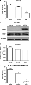
Fig. 1.
Fig. 1.
SBP1 inhibited GPX1 activity in HCT116 cells and MCF-7 cells but did not affect GPX1 protein and mRNA levels. (A) Endogenous GPX1 activity was inhibited by SBP1 in HCT116 cells. HCT116 parental (mock), pIRES2 empty vector control (pIRES2) and SBP1 overexpressing HCT116 cells (SBP1) were lysed and GPX1 activity determined (*P < 0.001) (n = 3, indicating the experiments were repeated three times). (B) GPX1 protein was not affected by SBP1 overexpression. The HCT116 parental, pIRES2 empty vector control (pIRES2) and SBP1 overexpressing HCT116 cells (SBP1) were lysed and immunoblotted to determine GPX1 and SBP1 protein levels. GPX1 signals were quantified by densitometry and normalized to β-actin, a loading control. (C) GPX1 mRNA levels were not affected by SBP1 overexpression. Quantitative real-time PCR was used to quantify GPX1 mRNA in HCT116 parental, pIRES2 control and SBP1 overexpressing HCT116 cells. The data were normalized to HCT116 parental cells (n = 3). (D) GPX1 activity derived from an expression construct was inhibited by SBP1 in MCF-7-GPX1 cells. MCF-7-GPX1 cells were treated with transfection reagents without plasmid (mock), pIRES2 control vector or pIRES2-SBP1 plasmid. Forty-eight hours after transfection, cells were harvested and GPX1 activity was determined (*P < 0.001) (n = 3).
Fig. 2.
Fig. 2.
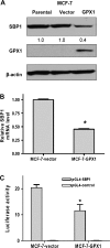
GPX1 negatively regulated SBP1 expression. (A) SBP1 protein was inhibited by GPX1. MCF-7 parental, MCF-7-vector and MCF-7-GPX1 stable transfected cells were lysed and subjected to immunoblotting for SBP1 and GPX1. SBP1 and GPX1 signals were quantified and normalized to β-actin, a loading control. Relative intensities were indicated under lanes. (B) SBP1 mRNA was significantly reduced in MCF-7-GPX1 cells compared with the MCF-7-vector cells (*P < 0.01) (n = 3). (C) GPX1 inhibited SBP1 promoter activity. MCF-7-vector and MCF-7-GPX1 cells were cotransfected with Renilla and the pGL4 control or pGL4-SBP1 promoter. Twenty-four hours after transfection, cells were harvested and luciferase activity determined (*P < 0.01). (n = 3). Each bar represents the mean ± SD of samples obtained in triplicate.
Fig. 3.
Fig. 3.
Contrasting effects of sodium selenite on SBP1 and GPX1. (A) Sodium selenite increased GPX1 expression and reduced SBP1 expression in the MCF-7-GPX1 cells with addition of 0, 25, 100 or 250 nM sodium selenite to the basal media, which contained at least 30 nM Se from the 10% fetal bovine serum for 48 h. Cells were harvested and immunoblotted to detect SBP1 and GPX1. SBP1 and GPX1 signals were quantified by densitometry and normalized to β-actin, a loading control, with the relative intensities indicated under the lanes. Similar experiments were performed in a HCT116-SBP1 stably transfected cell line. (B) SBP1 was not affected by sodium selenite in the MCF-7 cells that did not express GPX1. (C) All experiments were repeated at least three times and representative blots shown.
Fig. 4.
Fig. 4.
(A) SBP1 attenuated sodium selenite-induced increase of GPX1 activity in HCT116 cells. HCT116-pIRES2 control and HCT116-SBP1 stable cells were treated with addition of 0, 25, 100 or 250 nM sodium selenite to the basal media, which contained at least 30 nM Se from the 10% fetal bovine serum. Forty-eight hours after treatment, the cells were lysed and GPX1 activity was determined. (B) GPX1 levels increased and SBP1 declined with supplementation of sodium selenite, reaching a maximum at 250 nM and then the trends reversed with increasing supplementation with GPX1 declining and SBP1 increasing at the higher selenium concentrations. SBP1 and GPX1 signals were quantified by densitometry and normalized to β-actin, a loading control, with the relative intensities indicated under the lanes. All experiments were repeated at least three times and representative blots shown.
Fig. 5.
Fig. 5.
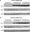
Dietary selenium status affected GPX1 and SBP1 expression in mouse colonic (A) and duodenal epithelial cells (B). Mice were fed with selenium-adequate diet (0.1 p.p.m.), selenium-deficient diet (0 p.p.m., Se-deficiency) or selenium-enriched diet (0.4 p.p.m. Se-supplemental) for 10 weeks. Intestinal epithelial cells were isolated for western blotting analysis for GPX1 and SBP1. β-actin was used as loading control. Each lane represented one individual mouse and three mice from each group were studied.
Fig. 6.
Fig. 6.
SBP1 interacts with GPX1 as determined by immunoprecipitation (IP). HCT116 cells were cotransfected with SBP1 expression plasmid pcDNA-HA-SBP1 and GPX1 expression plasmid pECFP-GPX1 or empty vector pECFP or Sec-Cys GPX1 plasmid (pECFP-GPX1-Cys). (A) Whole-cell lysates were analyzed by immunoblotting (IB) to confirm transfection efficiency and downregulation of SBP1 by GPX1. (B) The cell lysates were incubated with anti-GFP antibody to immunoprecipitate pECFP-GPX1 and pECFP-GPX1-Cys and then probed with anti-HA antibody to detect the HA-tagged SBP1. Non-specific peptide (NSP) in the same blot showed the equal immunoprecipitation of pECFP, pECFP-GPX1 and pECFP-GPX1-Cys. Lane 1, cell lysate from cells cotransfected with HA-SBP1 and pECFP; lane 2, cell lysate from cells cotransfected with HA-SBP1 and pECFP-GPX1 and lane 3, cell lysate from cells cotransfected with HA-SBP1 and pECFP-GPX1-Cys. Equal amount of protein were added to the gel and all experiments were repeated at least three times with representative blots shown.

Similar articles

Cited by

References

    1. Behne D, et al. Mammalian selenium-containing proteins. Annu. Rev. Nutr. 2001;21:453–473. - PubMed
    1. Kryukov GV, et al. Characterization of mammalian selenoproteomes. Science. 2003;300:1439–1443. - PubMed
    1. Hatfield DL, et al. How selenium has altered our understanding of the genetic code. Mol. Cell. Biol. 2002;22:3565–3576. - PMC - PubMed
    1. Wang L, et al. Methyl-selenium compounds inhibit prostate carcinogenesis in the transgenic adenocarcinoma of mouse prostate model with survival benefit. Cancer Prev. Res. (Phila Pa) 2009;2:484–495. - PMC - PubMed
    1. Clark LC, et al. Effects of selenium supplementation for cancer prevention in patients with carcinoma of the skin. A randomized controlled trial. Nutritional Prevention of Cancer Study Group. JAMA. 1996;276:1957–1963. - PubMed

Publication types

MeSH terms