Comparative Study
. 2010 Jul 1;185(1):507-16.
doi: 10.4049/jimmunol.0903598. Epub 2010 Jun 7.

An evaluation of the role of properdin in alternative pathway activation on Neisseria meningitidis and Neisseria gonorrhoeae

Affiliations
Comparative Study

An evaluation of the role of properdin in alternative pathway activation on Neisseria meningitidis and Neisseria gonorrhoeae

Sarika Agarwal et al. J Immunol. .

Abstract

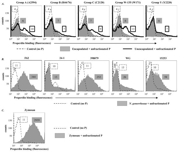
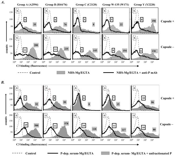
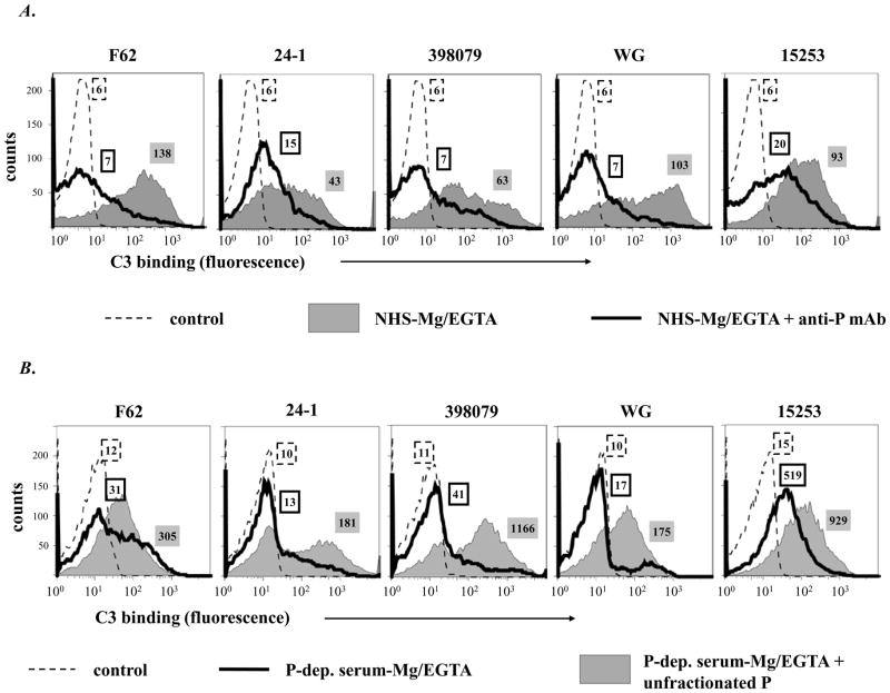
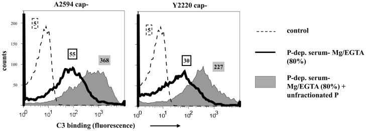
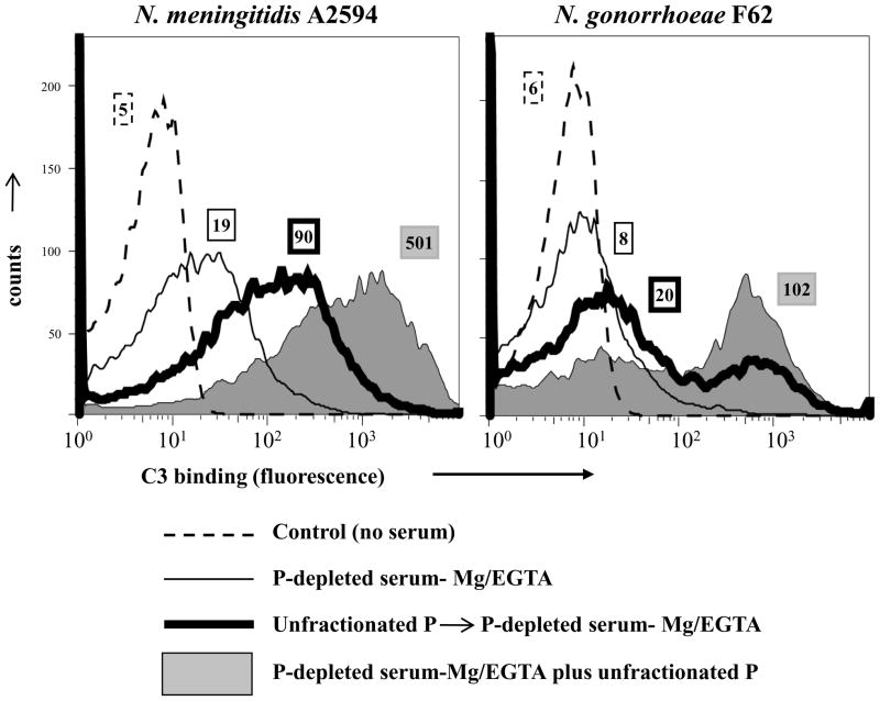
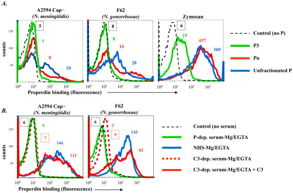
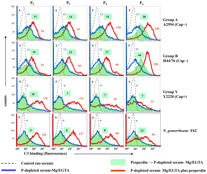
Properdin, a positive regulator of the alternative pathway (AP) of complement is important in innate immune defenses against invasive neisserial infections. Recently, commercially available unfractionated properdin was shown to bind to certain biological surfaces, including Neisseria gonorrhoeae, which facilitated C3 deposition. Unfractionated properdin contains aggregates or high-order oligomers, in addition to its physiological "native" (dimeric, trimeric, and tetrameric) forms. We examined the role of properdin in AP activation on diverse strains of Neisseria meningitidis and N. gonorrhoeae specifically using native versus unfractionated properdin. C3 deposition on Neisseria decreased markedly when properdin function was blocked using an anti-properdin mAb or when properdin was depleted from serum. Maximal AP-mediated C3 deposition on Neisseriae even at high (80%) serum concentrations required properdin. Consistent with prior observations, preincubation of bacteria with unfractionated properdin, followed by the addition of properdin-depleted serum resulted in higher C3 deposition than when bacteria were incubated with properdin-depleted serum alone. Unexpectedly, none of 10 Neisserial strains tested bound native properdin. Consistent with its inability to bind to Neisseriae, preincubating bacteria with native properdin followed by the addition of properdin-depleted serum did not cause detectable increases in C3 deposition. However, reconstituting properdin-depleted serum with native properdin a priori enhanced C3 deposition on all strains of Neisseria tested. In conclusion, the physiological forms of properdin do not bind directly to either N. meningitidis or N. gonorrhoeae but play a crucial role in augmenting AP-dependent C3 deposition on the bacteria through the "conventional" mechanism of stabilizing AP C3 convertases.


Figures

Figure 1
Figure 1
Binding of properdin to Neisseriae following incubation with commercially available unfractionated purified human properdin (10 μg/ml). A. Five encapsulated strains of N. meningitidis (one representative strain from each of the five major serogroups – A, B, C, W-135 and Y) and their isogenic unencapsulated mutants. Bacteria-bound properdin was detected by flow cytometry using an anti-properdin mAb (Quidel; Cat. No. A235) followed by anti-mouse IgG conjugated to Alexafluor 647. In all graphs, the x-axis represents fluorescence on a log10 scale and the y-axis the number of events. Controls, where properdin was omitted from the reaction mixture, are shown by the broken line histograms. Numbers adjacent to the histograms represent median fluorescence intensities (MFIs) of P binding; numbers in the box with the dashed border represents fluorescence of the control bacteria, numbers in the grey shaded box binding to encapsulated bacteria and the box with a solid black outline binding to unencapsulated meningococci. B. properdin binding to five strains of N. gonorrhoeae and C. properdin binding to zymosan. One experiment of two reproducibly repeated experiments is shown for each of the histograms.
Figure 2
Figure 2
Properdin augments AP-mediated C3 deposition on N. meningitidis. A. Five encapsulated strains of N. meningitidis (upper panel) and their isogenic unencapsulated mutants (lower panel) were incubated with NHS-Mg/EGTA (20% (v/v)) either in the absence (grey shaded histograms) or in the presence (histograms depicted by solid lines) of an anti-properdin mAb that blocks properdin function (Quidel; Cat. No. A233). Total C3 deposited on the bacterial surface was measured by flow cytometry using sheep polyclonal anti-human C3c conjugated to FITC. B. C3 deposition was assessed on the ten strains of N. meningitidis up on incubation with properdin (P-) depleted serum-Mg/EGTA (20% (v/v)) (histograms depicted by solid lines), or with P-depleted serum-Mg/EGTA reconstituted with purified unfractionated properdin to a concentration of 10 μg/ml (grey shaded histograms). Detection of C3 was performed as indicated above. Axes are as described in Figure 1. Controls (no serum added to bacteria) are shown by the broken lines. Controls with heat-inactivated serum show nearly identical tracings and have been omitted for simplicity. Numbers adjacent to histograms represent the median fluorescence intensities (MFIs) of C3 binding. One representative experiment of two separately performed and reproducibly repeated experiments is shown for each of the histograms.
Figure 3
Figure 3
Properdin augments AP-mediated C3 deposition on N. gonorrhoeae. A. C3 deposition on 5 strains of N. gonorrhoeae incubated with NHS-Mg/EGTA (20% (v/v) alone (grey shaded histograms) or with added anti-P mAb (Quidel mAb A233) that blocks properdin function (histograms depicted by solid lines). B. C3 deposition on gonococcal strains after incubation with P-depleted serum-Mg/EGTA (20% (v/v)) (histograms depicted by solid lines) or with P-depleted serum-Mg/EGTA reconstituted with purified unfractionated P (grey shaded histograms). Numbers adjacent to histograms represent the median fluorescence intensities (MFIs) of C3 binding. Detection of C3 fragments was performed as in Fig. 1A. One representative experiment of two separately performed and reproducibly repeated experiments is shown for each of the histograms..
Figure 4
Figure 4
Properdin (P) mediated deposition of C3 on Neisseriae at 80% serum concentration. Total C3 deposition on the unencapsulated derivatives of strains A2594 and Y2220 incubated either with P-depleted serum-Mg/EGTA (final concentration 80% (v/v); solid black histograms) or P-depleted serum-Mg/EGTA(final concentration 80% (v/v)) reconstituted with purified unfractionated properdin (10 μg/ml; shaded grey histograms). Numbers adjacent to histograms represent the median fluorescence intensities (MFIs) of C3 binding. Detection of C3 fragments was performed as in Fig. 1A. One representative experiment of two separately performed and reproducibly repeated experiments is shown for each of the histograms.
Figure 5
Figure 5
Preincubation of Neisseriae with unfractionated properdin (P) enhances C3 deposition. Unencapsulated mutant of meningococcal strain A2594 and N. gonorrhoeae strain F62 were incubated either with P-depleted serum-Mg/EGTA (thin solid lines) or preincubated with unfractionated P (10 μg/ml). Organisms were washed then P-depleted serum-Mg/EGTA (labeled ‘Unfractionated P→P-depleted serum-Mg/EGTA’) was added (thick black lines) or organisms were incubated with P-depleted serum-Mg/EGTA reconstituted with unfractionated P (shaded grey histograms). Numbers adjacent to histograms represent the median fluorescence intensities (MFIs) of C3 binding. Detection of C3 fragments was performed as in Fig. 1A. One representative experiment of two separately performed and reproducibly repeated experiments is shown for each of the histograms.
Figure 6
Figure 6
Binding of fractionated properdin (P) to Neisseriae. A. P was fractionated into dimers (P2), trimers (P3), tetramers (P4) and higher order oligomers (Pn; properdin in the void volume of a size-exclusion chromatograph). Binding of each of these fractions to N. meningitidis A2594 (unencapsulated) and N. gonorrhoeae F62 was measured by flow cytometry. Binding of P2, P3 and P4 yielded nearly identical results; results with the P3 fraction (only) are shown for simplicity (solid green line). Binding of Pn is shown by the red line; binding of commercially available unfractionated P by the blue line. B. Serum P binds to Neisseriae in the presence of the alternative pathway of complement. P binding to unencapsulated N. meningitidis A2594 and N. gonorrhoeae F62 following incubation with C3-depleted serum-Mg/EGTA (broken red histograms), P-depleted serum-Mg/EGTA(green histograms), C3-depleted serum-Mg/EGTA reconstituted with purified C3 (solid red histograms), or normal human serum-Mg/EGTA (blue histograms). All sera contained 10 mM Mg2+ and 10 mM EGTA and the final concentration of sera in all reaction mixtures was 20%. Axes are as described in Figure 1. Controls (no added serum) are shown by the black broken lines. Numbers adjacent to histograms represent the median fluorescence intensities (MFIs) of P binding. One representative experiment of two separately performed and reproducibly repeated experiments is shown for each of the histograms.
Figure 7
Figure 7
Preincubation with native properdin (P) does not enhance C3 deposition on Neisseriae. Unencapsulated derivatives of serogroup A strain 2594 and serogroup B strain H44/76, encapsulated serogroup Y strain 2220 and gonococcal strain F62 were pre-incubated either with properdin dimers (P2), trimers (P3), tetramers (P4) or higher-order oligomers (Pn) each to a final concentration of 10 μg/ml and washed. P-depleted serum-Mg/EGTA was then added to a final concentration of 20%. C3 deposition on bacteria was measured by flow cytometry (shaded green histograms). Bacteria incubated only with P-depleted serum-Mg/EGTA (final concentration 20%) are shown by the blue histograms. C3 deposition was also measured on bacteria that were incubated with P--depleted serum-Mg/EGTA that had been reconstituted with each of the purified properdin (P2, P3, P4 or higher-order oligomer [Pn]) fractions (final concentration of each P fraction was 10 μg/ml; shown by the red histograms). Axes are as described in Figure 1. Controls (serum lacking from the reaction mixture) are indicated by the broken lines. Numbers adjacent to histograms represent the median fluorescence intensities (MFIs) of P binding. One representative of two separately performed and reproducibly repeated experiments is shown for each of the histograms.

References

    1. Nolan KF, Kaluz S, Higgins JM, Goundis D, Reid KB. Characterization of the human properdin gene. Biochem J. 1992;287(Pt 1):291–297. - PMC - PubMed
    1. Pangburn MK. Analysis of the natural polymeric forms of human properdin and their functions in complement activation. J Immunol. 1989;142:202–207. - PubMed
    1. Smith CA, Pangburn MK, Vogel CW, Muller-Eberhard HJ. Molecular architecture of human properdin, a positive regulator of the alternative pathway of complement. JBiol Chem. 1984;259:4582–4588. - PubMed
    1. Fearon DT, Austen KF. Properdin: binding to C3b and stabilization of the C3b-dependent C3 convertase. J Exp Med. 1975;142:856–863. - PMC - PubMed
    1. Pillemer L, Blum L, Lepow IH, Ross OA, Todd EW, Wardlaw AC. The properdin system and immunity. I. Demonstration and isolation of a new serum protein, properdin, and its role in immune phenomena. Science. 1954;120:279–285. - PubMed

Publication types

MeSH terms