. 2010 Jul 1;185(1):367-75.
doi: 10.4049/jimmunol.0903627. Epub 2010 Jun 7.

Asymmetric cell division of T cells upon antigen presentation uses multiple conserved mechanisms

Affiliations

Asymmetric cell division of T cells upon antigen presentation uses multiple conserved mechanisms

Jane Oliaro et al. J Immunol. .

Abstract

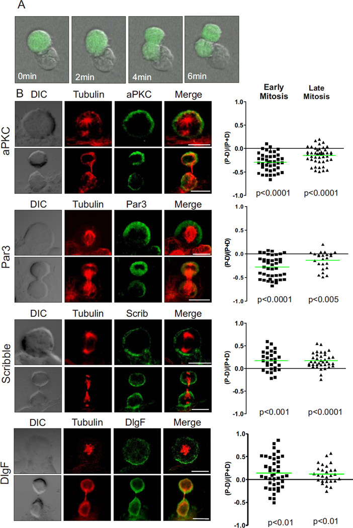
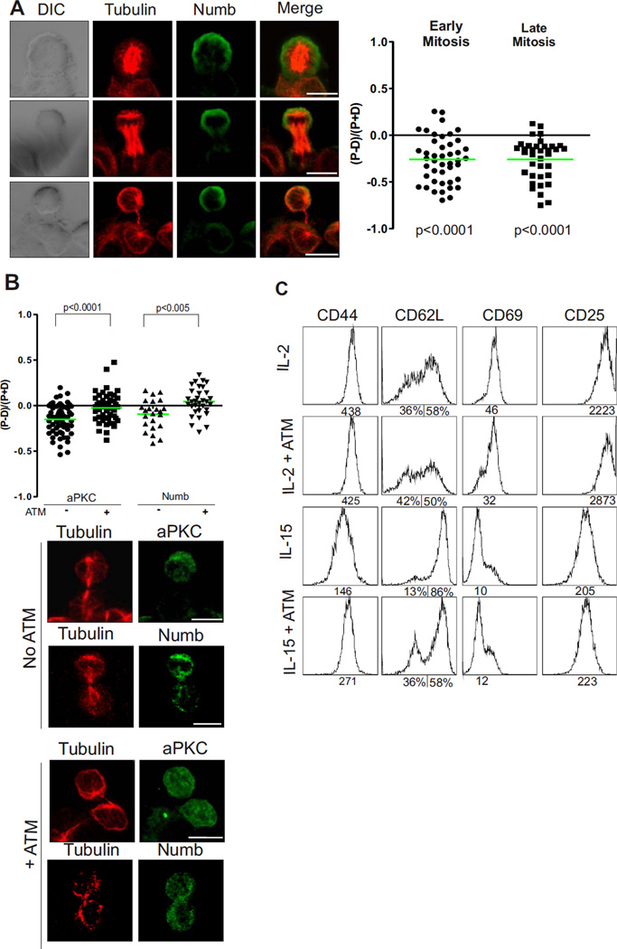
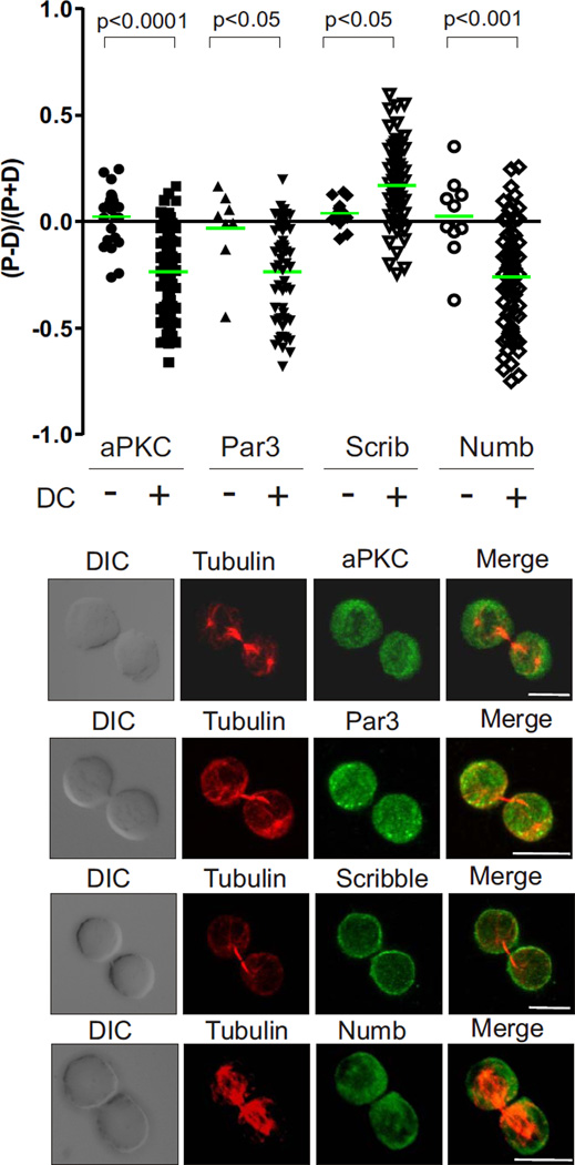
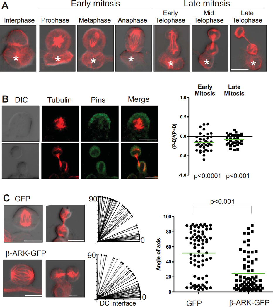
Asymmetric cell division is a potential means by which cell fate choices during an immune response are orchestrated. Defining the molecular mechanisms that underlie asymmetric division of T cells is paramount for determining the role of this process in the generation of effector and memory T cell subsets. In other cell types, asymmetric cell division is regulated by conserved polarity protein complexes that control the localization of cell fate determinants and spindle orientation during division. We have developed a tractable, in vitro model of naive CD8(+) T cells undergoing initial division while attached to dendritic cells during Ag presentation to investigate whether similar mechanisms might regulate asymmetric division of T cells. Using this system, we show that direct interactions with APCs provide the cue for polarization of T cells. Interestingly, the immunological synapse disseminates before division even though the T cells retain contact with the APC. The cue from the APC is translated into polarization of cell fate determinants via the polarity network of the Par3 and Scribble complexes, and orientation of the mitotic spindle during division is orchestrated by the partner of inscuteable/G protein complex. These findings suggest that T cells have selectively adapted a number of evolutionarily conserved mechanisms to generate diversity through asymmetric cell division.


Figures

Figure 1
Figure 1
Polarity proteins are asymmetric in mitotic T cells interacting with DC. (A) Naïve OT-1 CD8+ T cells expressing GFP were cultured with peptide-pulsed dendritic cells. The co-cultures were left for 40 hrs prior to imaging and then imaged by time-lapse microscopy every two minutes. Still images show an example of a T cell dividing while attached to a dendritic cell - time stamp shows time of progression through mitosis. Scale bar: 25 µm. (B) Naïve OT-1 CD8+ T cells dividing in response to antigen presentation by DC were fixed and stained to determine the ratio of proximal to distal polarization of aPKC, PAR-3, Scribble, and DlgF (3 experiments; 73, 61, 66 and 80 cells were analyzed respectively, representative images on the left). Positive and negative values indicate proximal and distal polarization respectively. Each point on the graph represents an individual cell and the green bar represents the mean. Scale bars: 10 µm.
Figure 2
Figure 2
The polarity network orchestrates asymmetric distribution of the cell fate determinant, Numb. (A) The ratio of proximal to distal polarization was assessed as in Fig 2 for Numb in different phases of mitosis (3 experiments; 80 cells). Representative images on left, scale bars: 10 µm. (B) The ratio of proximal to distal polarization was assessed as in Fig 2 for aPKC (61 & 50 cells) and Numb (57 & 66 cells) with or without treatment with ATM (2 experiments). Representative images below, scale bars: 10 µm, NS = not significant. (C) Expression of the surface markers CD44, CD62L, CD69, CD25 on OT-1 CD8+ T cells cultured under effector (IL-2) and memory (IL-15) conditions with or without treatment with ATM. The geometric mean of the individual peaks, or % cells gated, is shown under the plots. Data is representative of 3 experiments.
Figure 3
Figure 3
ACD of T cells requires contact with the antigen presenting cell. The ratio of proximal to distal polarization was assessed as in Fig 2 for aPKC (24 cells), Scribble (14 cells), PAR-3 (8 cells) and Numb (10 cells) in mitotic cells unattached to a DC (representative images below) compared to mitotic cells attached to a DC. Scale bars; 10 µm.
Figure 4
Figure 4
ACD of T cells requires contact with the DC, but not a sustained immunological synapse. The ratio of proximal to distal polarization was assessed as in Fig 2 for synapse and distal pole proteins CD8 (40 cells), LFA-1 (66 cells), PKCθ (79 cells) and CD45 (58 cells) (3 experiments). Representative images on left. Scale bars; 10 µm.
Figure 5
Figure 5
ACD of T cells utilizes the Pins/G protein module to orient the spindle. OT-1 CD8+ T cells dividing whilst attached to a DC were (A) stained for tubulin and scored for orientation of the MTOC or (B) stained for tubulin and Pins to assess the ratio of proximal or distal polarization (3 experiments; 73 cells) or (C) Naive OT-T cells expressing GFP (n=75) or β-ARK-GFP (n=70) were incubated with DC and stained for tubulin to mark T cells in division. Image J software was used to draw a line through the axis of division based on the position of the centrosomes, using the DC interface as the horizontal axis and used to calculate the angle of spindle axis relative to the DC interface and plotted on the right (2 experiments, representative images on left). Scale bars; 10 µm.

Similar articles

Cited by

References

    1. Williams MA, Bevan MJ. Effector and memory CTL differentiation. Annu Rev Immunol. 2007;25:171–192. - PubMed
    1. Kaech SM, Wherry EJ. Heterogeneity and cell-fate decisions in effector and memory CD8+ T cell differentiation during viral infection. Immunity. 2007;27:393–405. - PMC - PubMed
    1. Russell SM. Determination of T-cell fate by dendritic cells: a new role for asymmetric cell division? Immunol Cell Biol. 2008;86:423–427. - PubMed
    1. Chang JT, Palanivel VR, Kinjyo I, Schambach F, Intlekofer AM, Banerjee A, Longworth SA, Vinup KE, Mrass P, Oliaro J, Killeen N, Orange JS, Russell SM, Weninger W, Reiner SL. Asymmetric T lymphocyte division in the initiation of adaptive immune responses. Science. 2007;315:1687–1691. - PubMed
    1. Reiner SL, Sallusto F, Lanzavecchia A. Division of labor with a workforce of one: challenges in specifying effector and memory T cell fate. Science. 2007;317:622–625. - PubMed

Publication types

MeSH terms