. 2010 Aug 13;285(33):25582-90.
doi: 10.1074/jbc.M110.133215. Epub 2010 Jun 8.

Structural insights into inhibition of sterol 14alpha-demethylase in the human pathogen Trypanosoma cruzi

Affiliations

Structural insights into inhibition of sterol 14alpha-demethylase in the human pathogen Trypanosoma cruzi

Galina I Lepesheva et al. J Biol Chem. .

Abstract

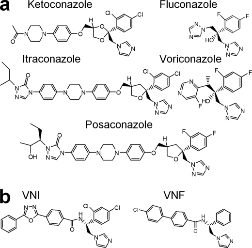
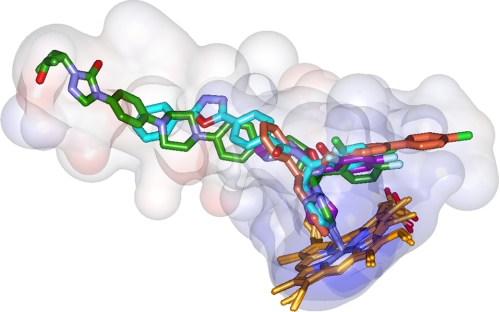
Trypanosoma cruzi causes Chagas disease (American trypanosomiasis), which threatens the lives of millions of people and remains incurable in its chronic stage. The antifungal drug posaconazole that blocks sterol biosynthesis in the parasite is the only compound entering clinical trials for the chronic form of this infection. Crystal structures of the drug target enzyme, Trypanosoma cruzi sterol 14alpha-demethylase (CYP51), complexed with posaconazole, another antifungal agent fluconazole and an experimental inhibitor, (R)-4'-chloro-N-(1-(2,4-dichlorophenyl)-2-(1H-imid-azol-1-yl)ethyl)biphenyl-4-carboxamide (VNF), allow prediction of important chemical features that enhance the drug potencies. Combined with comparative analysis of inhibitor binding parameters, influence on the catalytic activity of the trypanosomal enzyme and its human counterpart, and their cellular effects at different stages of the Trypanosoma cruzi life cycle, the structural data provide a molecular background to CYP51 inhibition and azole resistance and enlighten the path for directed design of new, more potent and selective drugs to develop an efficient treatment for Chagas disease.


Figures

FIGURE 1.
FIGURE 1.
14DM inhibitors. a, five azoles currently available for systemic use as clinical antifungal drugs. b, VNI and VNF represent a novel experimentally identified inhibitory scaffold selective for trypanosomal 14DMs.
FIGURE 2.
FIGURE 2.
Overall 14DM structure. Posaconazole-bound TC14DM (rainbow color, from blue N terminus to red C terminus) is superimposed with the ligand-free Tbb14DM (semitransparent gray); root mean square deviation for all Cα atoms is 0.91 Å. Major P450 structural elements are marked. The heme is shown in stick representation; posaconazole was deleted for clarity. The most flexible areas (GH loop and FG loop regions) are marked with orange arrows, 1 and 2, respectively. Repositioning the rigid I helix that is observed in all inhibitor bound trypanosomal 14DMs is marked with a blue arrow 3.
FIGURE 3.
FIGURE 3.
Location of the inhibitors in the TC14DM structures (left) and stereoview of the active site (right) in complexes with: posaconazole (a), fluconazole (b), and VNF (c). The protein is in the same orientation shown from the distal view; the backbone is displayed as gray ribbons. The active site surfaces (Accelrys) are colored by interpolating charge; the inhibitors and the heme are shown in stick representation; the residues located within Van der Waals bond distances (4.5 Å) are depicted by lines. For posaconazole, fluconazole, and VNF (throughout all figures) the carbons are colored in green, violet, and salmon, respectively; the heme carbons are orange. Nitrogens are blue, oxygens are red, chlorines are gray, fluorines are light blue, sulfurs are orange; protein atoms follow the same scheme except carbons are gray. Residue labels are color-coded according to their specificity, with green labels for the posaconazole and salmon labels for VNF-specific residues. The list of inhibitor-contacting residues in the three TC14DM structures and in the VNI-bound Tbb14DM is provided as supplemental Table S2. The electron density maps for the inhibitors are presented in supplemental Fig. S2.
FIGURE 4.
FIGURE 4.
Specific features in the binding of inhibitors. a, the long arm of posaconazole is extended above the protein surface forming 12 additional interactions around the access channel entrance (left panel). The surface is colored by hydrophobicity (from blue for the most polar to white to orange red for the most hydrophobic residues; the surfaces for the three TC14DM structures can be seen as supplemental Fig. S3). Right panel, posaconazole in the TC14DM structure (stick representation) superimposed with its energy-minimized model (line representation). Location of Gly49 in the structure and models of its mutations to the residues found in the posaconazole-resistant A. fumigatus 14DM are shown. b, fluconazole alters positions of conserved Tyr103 and Tyr116 affecting their hydrogen bonding with the heme propionates (rings A and D). The corresponding tyrosines and the heme in the superimposed posaconazole bound TC14DM structure are shown as green lines; the hydrogen bonds are depicted as dotted lines. c, VNF (salmon) binds to TC14DM in the orientation opposite to that of its scaffold analog VNI (cyan) in the Tbb14DM structure. Two water molecules (Tbb14DM) are displayed as red spheres (left panel). Right panel, the longer arm of VNF is intercalated between the B′ helix/B′C loop, helix C, and N-terminal (N-term) part of helix I, slightly broadening the active site cavity. The corresponding structural elements in the superimposed posaconazole-bound TC14DM are colored in green.
FIGURE 5.
FIGURE 5.
14DM expression, cellular localization, and inhibition in T. cruzi. a, immunoblot analysis of 14DM expression in different life stages of the parasite. Equal concentrations of whole cell extracts of trypomastigotes (T), epimastigotes (E), and amastigotes (A) were separated by SDS-PAGE, blotted onto nitrocellulose, probed with mouse anti-Tbb14DM antiserum, and densitometrically scanned. The values (indicated by an asterisk) correspond to relative intensities normalized to the β-actin loading control. b, localization of 14DM in T. cruzi by immunogold electron microscopy. 14DM (arrows) is seen in the cytoplasm and reservosomes (R). N, nucleus. c, inhibition of the ability of trypomastigotes to infect cardiomyocytes by fluconazole (Fluc), posaconazole (Pos), and VNF. GFP-expressing trypomastigotes were pretreated with 14DM inhibitors and exposed to cardiomyocytes monolayers, and infected monolayers were incubated for 72 h to assay for parasite multiplication. The infection was evaluated by GFP fluorescence (RFU). The data represent the mean ± S.D. of the results from triplicate samples. d, fluorescence microscopic observation of the inhibition of T. cruzi multiplication by 1 μm 14DM inhibitors within cardiomyocytes. GFP-expressing amastigotes are seen as small green spots inside cardiomyocytes, and cardiomyocytes nuclei are blue. e, 14DM expression upon exposure of T. cruzi to azole inhibitors. Epimastigotes (106 organisms/ml) were allowed to grow for 5 days after exposure to inhibitors. 14DM expression was analyzed as in a, the values (indicated by an asterisk) were normalized to α-tubulin. f, dose-dependent inhibition of T. cruzi epimastigotes multiplication by 14DM inhibitors. The data represent the mean ± S.D. of the results from triplicate samples.
FIGURE 6.
FIGURE 6.
14DM structure-based inhibitory pharmacophore. Posaconazole is green, fluconazole is violet, VNF is salmon (TC14DM), and VNI (Tbb14DM) is cyan. Semitransparent (4-Å radius) surfaces are shown. Stereoview of the active site including contacting residues from all four structures can be seen in supplemental Fig. S5.

Similar articles

Cited by

References

    1. Ekroos M., Sjogren T. (2006) Proc. Natl. Acad. Sci. U.S.A. 103, 13682–13687 - PMC - PubMed
    1. Moncayo A., Silveira A. C. (2009) Mem. Inst. Oswaldo Cruz. 104, 17–30 - PubMed
    1. Neva F. A. (2007) Cecil Medicine:Expert Consult, Ch. 368, 23rd Ed., Saunders Elsevier, Philadelphia
    1. Schmunis G. A. (2007) Mem. Inst. Oswaldo Cruz. 102, 75–85 - PubMed
    1. Gonzalez-Granado L. I., Rojo-Conejo P., Ruiz-Contreras J., Gonzalez-Tomé M. I. (2009) Lancet 373, 2025. - PubMed

Publication types

MeSH terms

LinkOut - more resources