. 2010 Oct;38(19):6673-83.
doi: 10.1093/nar/gkq501. Epub 2010 Jun 8.

The GW/WG repeats of Drosophila GW182 function as effector motifs for miRNA-mediated repression

Affiliations

The GW/WG repeats of Drosophila GW182 function as effector motifs for miRNA-mediated repression

Marina Chekulaeva et al. Nucleic Acids Res. 2010 Oct.

Abstract

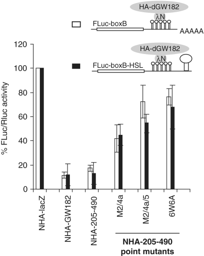
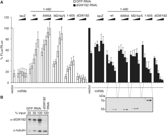
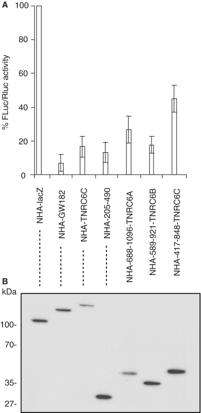
The control of messenger RNA (mRNA) function by micro RNAs (miRNAs) in animal cells requires the GW182 protein. GW182 is recruited to the miRNA repression complex via interaction with Argonaute protein, and functions downstream to repress protein synthesis. Interaction with Argonaute is mediated by GW/WG repeats, which are conserved in many Argonaute-binding proteins involved in RNA interference and miRNA silencing, from fission yeast to mammals. GW182 contains at least three effector domains that function to repress target mRNA. Here, we analyze the functions of the N-terminal GW182 domain in repression and Argonaute1 binding, using tethering and immunoprecipitation assays in Drosophila cultured cells. We demonstrate that its function in repression requires intact GW/WG repeats, but does not involve interaction with the Argonaute1 protein, and is independent of the mRNA polyadenylation status. These results demonstrate a novel role for the GW/WG repeats as effector motifs in miRNA-mediated repression.


Figures

Figure 1.
Figure 1.
205–490 region of dGW182 is the minimal N-terminal effector domain (NED). (A) Schematic representation of Drosophila GW182 protein and its deletion mutants. The numbers correspond to the amino acid positions. (B) Schematic representation of reporter constructs: FLuc-boxB contains firefly luciferase (FLuc) coding sequence and 3′UTR with five boxB sites specifically binding to λN peptide; RLuc contains Renilla luciferase (RLuc) coding sequence and no boxB sites (34). (C) Repression of FLuc-boxB mRNA by NHA-dGW182 and its deletion mutants. Drosophila S2 cells were co-transfected with plasmids encoding FLuc-boxB, RLuc, and full-length NHA-dGW182 or indicated NHA-dGW182 deletion mutants. As negative controls, plasmids encoding either NHA alone or NHA fused to β-galactosidase (NHA-lacZ) were used. Expression of FLuc was normalized to that of RLuc. Values are presented as a percentage of FLuc produced in the presence of NHA-lacZ. Values represent means ±SD from at least four experiments. (D) Expression of different HA-fusion proteins was estimated by western blotting with antibodies directed against HA-peptide.
Figure 2.
Figure 2.
The N-terminal effector domain (NED) does not interact with AGO1. Drosophila S2 cells were transfected with plasmids encoding the full-length NHA-dGW182 or its deletion mutants. Cell lysates were used in immunoprecipitations with anti-AGO1 antibody. Inputs and immunoprecipitates were analyzed by western blotting using anti-HA or anti-AGO1 antibodies, as indicated on the left.
Figure 3.
Figure 3.
Mutation of the conserved amino acid residues abolish repression by NED. (A) Schematic representation of the NED region and its alignment across the species (Dm: Drosophila melanogaster, Da: Drosophila ananassae; Ap: Apis mellifera; TNRC6A, B, and C are human GW182 homologs). Alignment was performed with a T-Coffee tool [(41) see Supplementary Figure S1 for full alignment]. Mutated residues (always to alanine) are shown in red. The numbers correspond to amino acid position. Asterisks mark residues identical in all sequences, colons mark conservative substitutions, dots mark semi-conservative substitutions. (B) Repression of FLuc-boxB mRNA by the NED and its point mutants. Drosophila S2 cells were co-transfected with plasmids encoding FLuc-boxB, RLuc, and either NHA-205-490 (NED) or one of its point mutants depicted in (A). For other details, see legend to Figure 1. (C) Expression levels of HA-fusion proteins as estimated by western blotting with the anti-HA antibody.
Figure 4.
Figure 4.
Repression by the NED requires GW/WG repeats and involves mRNA destabilization. (A) NHA-205–490, either wild-type or mutants in which one or several GW/WG repeats are mutated (always W to A), were co-transfected with FLuc-boxB and RLuc. For other details, see Figure 1. (B) The levels of HA-fusion proteins as analyzed by western blotting with the anti-HA antibody. (C) The FLuc-boxB and RLuc mRNA levels from experiments depicted in Figures 4A and 5A were analyzed by northern blot. mRNA levels were quantified by phosphoimaging; amounts of FLuc-boxB were normalized according to Renilla control and expressed as a percentage of FLuc-boxB level in the presence of NHA-lacZ protein (numbers below the figure). Lanes 1–7 show the FLuc-boxB and RLuc RNA levels in the presence of non-tethered control HA-dGW182, tethered NHA-lacZ, NHA-dGW182, NHA-NED and its point mutants: M2/4a, M2/4a/5, and 6W6A, accordingly.
Figure 5.
Figure 5.
Repression mediated by the NED is independent of the poly(A) tail. Drosophila S2 cells were transfected with the boxB-containing FLuc reporters, either FLuc-boxB (open bars) or FLuc-boxB-HSL (black bars). Unlike FLuc-boxB, FLuc-boxB-HSL lacks a polyadenylation signal and bears instead a histone H1 stem-loop (HSL) structure in its 3′UTR. FLuc reporters were co-transfected with RLuc as a normalization control, and plasmids encoding indicated NHA-fusion proteins.
Figure 6.
Figure 6.
Overexpression of the NED fused to the AGO1-binding motif rescues RNAi knockdown of endogenous dGW182. (A) Endogenous dGW182 was depleted in Drosophila S2 cells with dsRNA (black bars); a batch of cells was treated with GFP-specific dsRNA as a negative control (open bars). Cells were transfected with firefly luciferase reporter containing miRNA-9b target sites (FLuc-nerfin) and with either the miRNA-9b-encoding plasmid or the empty vector. As in all transfection experiments, RLuc was co-expressed as a transfection control; expression levels of firefly luciferase were normalized to Renilla luciferase activity and expressed as a percentage of the firefly luciferase activity in the presence of the empty vector. To rescue the knockdown of endogenous dGW182, increasing amounts of plasmids encoding NHA-dGW182 or its N-terminal fragments were co-transfected: the NED fused to the AGO1-binding motif (1–490), either wild-type or non-functional point mutants (M2/4a/5 and 6W6A), and the 1–605 construct. Expression of NHA-dGW182 fragments was estimated by western blotting with anti-HA antibody. Increasing amounts of NHA-lacZ were co-transfected for a negative control. (B) Efficiency of the endogenous dGW182 knockdown was estimated by western blot analysis. S2 cells were treated with dsRNAs, indicated above the lanes (dGW182 or GFP as a negative control), and analyzed by western blotting with the antibodies shown on the left. Expression of tubulin was estimated as a loading control.
Figure 7.
Figure 7.
N-terminal domains of human GW182 homologs, TNRC6A, B and C, are able to repress tethered mRNA in Drosophila S2 cells. The assay was performed as in Figure 1. (A) The N-terminal fragments of human TNRC6A (Q8NDV7; amino acids 688–1096), TNRC6B (Q9UPQ9, amino acids 589–921), and TNRC6C (Q9HCJ0, amino acids 417–848) aligning with the Drosophila NED and enriched in GW-rich repeats (see Figure 3 and Supplementary Figure S1) were tethered to the FLuc-boxB reporter. dGW182 was tethered as a positive control, NHA-lacZ as the negative control. (B) Expression levels of NHA-fusion proteins were estimated by western blotting with anti-HA antibody.

Similar articles

Cited by

References

    1. Friedman RC, Farh KK, Burge CB, Bartel DP. Most mammalian mRNAs are conserved targets of microRNAs. Genome Res. 2009;19:92–105. - PMC - PubMed
    1. Eulalio A, Huntzinger E, Izaurralde E. Getting to the root of miRNA-mediated gene silencing. Cell. 2008;132:9–14. - PubMed
    1. Filipowicz W, Bhattacharyya SN, Sonenberg N. Mechanisms of post-transcriptional regulation by microRNAs: are the answers in sight? Nat. Rev. Genet. 2008;9:102–114. - PubMed
    1. Chekulaeva M, Filipowicz W. Mechanisms of miRNA-mediated post-transcriptional regulation in animal cells. Curr. Opin. Cell. Biol. 2009;21:452–460. - PubMed
    1. Nilsen TW. Mechanisms of microRNA-mediated gene regulation in animal cells. Trends Genet. 2007;23:243–249. - PubMed

Publication types