. 2010 Jul;137(13):2167-75.
doi: 10.1242/dev.051573.

Mesoderm migration in Drosophila is a multi-step process requiring FGF signaling and integrin activity

Affiliations

Mesoderm migration in Drosophila is a multi-step process requiring FGF signaling and integrin activity

Amy McMahon et al. Development. 2010 Jul.

Abstract

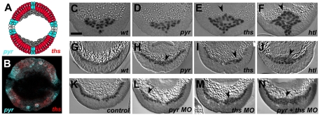
Migration is a complex, dynamic process that has largely been studied using qualitative or static approaches. As technology has improved, we can now take quantitative approaches towards understanding cell migration using in vivo imaging and tracking analyses. In this manner, we have established a four-step model of mesoderm migration during Drosophila gastrulation: (I) mesodermal tube formation, (II) collapse of the mesoderm, (III) dorsal migration and spreading and (IV) monolayer formation. Our data provide evidence that these steps are temporally distinct and that each might require different chemical inputs. To support this, we analyzed the role of fibroblast growth factor (FGF) signaling, in particular the function of two Drosophila FGF ligands, Pyramus and Thisbe, during mesoderm migration. We determined that FGF signaling through both ligands controls movements in the radial direction. Thisbe is required for the initial collapse of the mesoderm onto the ectoderm, whereas both Pyramus and Thisbe are required for monolayer formation. In addition, we uncovered that the GTPase Rap1 regulates radial movement of cells and localization of the beta-integrin subunit, Myospheroid, which is also required for monolayer formation. Our analyses suggest that distinct signals influence particular movements, as we found that FGF signaling is involved in controlling collapse and monolayer formation but not dorsal movement, whereas integrins are required to support monolayer formation only and not earlier movements. Our work demonstrates that complex cell migration is not necessarily a fluid process, but suggests instead that different types of movements are directed by distinct inputs in a stepwise manner.


Figures

Fig. 1.
Fig. 1.
Mesoderm migration is a multi-step process involving temporally distinct movements. (A,B,D,E,G,H) Embryo cross-sections stained with Twist antibody (black) to mark the mesoderm. Each stage is shown to demonstrate movement of the mesoderm over time: (A) stage 6, (B) stage 7, (D) stage 8, (E,G) stage 9 and (H) stage 10. Onset of germband elongation is represented by 0 minutes. Scale bar: 20 μm. (C) Collapse involves movement of mesoderm cells toward the ectoderm. Movement of mesoderm cells toward the ectoderm is represented by the radial axis of a cylinder, r (y-axis: 0=center of embryo, 90=ectoderm). The collapse of the mesoderm is shown as r over time, with each line representing movement of a single cell. Red is used to highlight the time period of collapse. (F) Spreading occurs after collapse and involves mesoderm cells crawling along the ectoderm, which is represented by the curvature of a cylinder, θ. Spreading is demonstrated by graphing θ over time (midline=0; dorsalmost points coincident with angular positions=1, –1). The timing of spreading is highlighted in blue. (I) Monolayer formation occurs last and involves incorporation of all cells into one layer via intercalation (see Fig. 5 for more details). Monolayer formation happens in the r direction from 75 minutes onward (highlighted in red).
Fig. 2.
Fig. 2.
pyr and ths mutants have a non-monolayer mesoderm phenotype. (A,B,G-N) Embryo cross-sections at stage 10. (C-F) Embryo cross-sections at stage 7. (A) Schematic of Pyr (blue) and Ths (red) expression in the ectoderm during mesoderm spreading. The receptor Htl is found in the mesoderm (gray). (B) Expression patterns of pyr (blue) and ths (red) transcript during mesoderm spreading detected by in situ hybridization. (C-J) Embryos of indicated genetic backgrounds sectioned and stained with anti-Twist antibody (black) in wild-type (C,G), pyre02915 (D,H), thse02026/thsDf238 (E,I) and htlAB42 (F,J) mutants. Arrowheads highlight defects. Morpholinos (MOs) were injected for live imaging purposes (see Materials and methods). Injection of gal4 MO (K), which does not have a target in Drosophila, did not affect mesoderm spreading, whereas injection of pyr MO (L) and ths MO (M) produced phenotypes similar to the genetic mutants. (N) Injection of pyr and ths MO together produced a phenotype similar to htl mutants. Scale bar: 20 μm.
Fig. 3.
Fig. 3.
Live imaging of FGF mutants using two-photon microscopy. Virtual cross-sections of H2A-GFP-expressing embryos taken from 4D imaging data sets (3D plus time) obtained on a two-photon microscope (see Materials and methods for details). (A) Wild-type embryos undergo characteristic movements: invagination at stage 6, collapse of the mesodermal tube at stage 7, spreading at stage 8 and 9 and monolayer formation at stage 10. (B) htl mutant embryo at stages 6-10. htl mutants have a collapse defect at stage 7 and a severe non-monolayer at stage 10. (C) pyr mutant embryo at stages 6-10. pyr mutant embryos undergo normal collapse and spreading during stages 6-9. A subset of cells are observed outside the monolayer at stage 10 (arrowhead). (D) ths mutant embryo at stages 6-10. In ths mutants, collapse is defective at stage 7 and a severe non-monolayer is observed at stage 10. Scale bar: 20 μm.
Fig. 4.
Fig. 4.
Live imaging and nuclear tracking reveals defects in ths mutants. (A) Drosophila embryos are roughly cylindrically shaped such that movement of mesoderm cells along the dorsoventral axis can be represented by the curve of a cylinder, θ (0=midline). Movement along the radial axis r represents movement of mesoderm cells toward or away from the ectoderm (0=center of embryo). (B) A color code is applied to track the progress of each cell over time, with a color assignment given at stage 6 and retained throughout migration. The color code is along the radial axis, where red represents mesoderm cells closest to the ectoderm at stage 6 and blue represents the furthest mesoderm cells. (C,E,G,I) Collapse of the mesodermal tube as shown by a graph of r over time; each curve represents the movement of one cell (y-axis: 0=center of embryo, 90=ectoderm; the black line is the average of all tracks). White boxes highlight the time intervals of collapse and intercalation in wild-type embryos defined in Fig. 1. (C) Wild-type embryos undergo collapse of the mesodermal tube to flatten along the ectoderm. Mesoderm cells in htl mutants (E) and ths mutants (G) fail to collapse. (I) pyr mutants display no collapse defect. (D,F,H,J) Spreading of mesoderm cells away from the midline (0) toward the dorsalmost point of the embryo (1 or –1) is shown by graphs of θ over time. The black line is the average of all tracks. White boxes highlight the time intervals of spreading in wild-type embryos as defined in Fig. 1. (D) Wild-type mesoderm cells spread directionally away from the midline toward the dorsal-most point in the embryo, whereas htl mutants (F) have aberrant spreading behavior, with some cells crossing over the midline and spreading in the wrong direction. ths (H) and pyr (J) mutants spread directionally away from the midline toward dorsal regions.
Fig. 5.
Fig. 5.
Intercalation of mesoderm cells during monolayer formation is disrupted in FGF mutants. (A) Intercalation occurs during mesoderm migration when a cell that is not in contact with the ectoderm (blue) moves toward the ectoderm. (B-I) A subset of mesoderm cells are tracked from stage 9 (B,D,F,H) to 10 (C,E,G,I) (gray ball=mesoderm cell), showing how cells go from a multilayer to a monolayer in wild-type embryos (B,C) but not in pyr (D,E), ths (F,G) or htl (H,I) mutants. Arrowheads demonstrate cells that have not intercalated. The view shown is similar to a cross-section as in Fig. 2. (J) A graph of stable intercalation of mesoderm cells over time. The number of cells that intercalate stably into the monolayer is highest for wild-type embryos, whereas pyr, ths and htl mutants have successively lower numbers of intercalating cells. The differences between pairs of phenotypes are all statistically significant (P<0.002). Scale bar: 20 μm.
Fig 6.
Fig 6.
Rap1 and Mys are required for monolayer formation. (A-H) Cross sections of embryos stained with Twist antibody (black). (A-D) Stage 7 embryos and (E-H) stage 10 embryos. (A,E) Wild-type embryos undergo tube collapse at stage 7 (A) and then intercalation to form a monolayer during stage 10 (E). (B-D,F-H) In htl mutants (B) and Rap1 mutants (C), tube collapse is defective, resulting in a clump of cells at stage 7. Intercalation is also affected, resulting in the lump remaining at stage 10 (F,G). In mys mutants, tube collapse is normal, resulting in normal mesoderm behavior at stage 7 (D). During stage 10, a non-monolayer is observed (H, arrowheads). (I-K) Cross-sections of embryos at stage 10 stained with Mys antibody (black). (I) In wild-type embryos, Mys is expressed at the boundary between the mesoderm and ectoderm. (J) In htl mutants, Mys levels are reduced and gaps in expression are observed (arrows). (K) Rap1 mutant embryos fail to localize Mys at the ectoderm-mesoderm boundary. Scale bars: 20 μm.
Fig. 7.
Fig. 7.
Mys is required for monolayer formation and mesoderm cell shape changes. (A,B) Collapse and spreading of mesoderm cells in mys mutants represented by r and θ over time, respectively (see Fig. 4 for more details). A radial color code is applied to distinguish each cell track over time. The black line represents the average behavior of all mesoderm cells. (C) Monolayer formation is measured as the percent of cells that are incorporated by stable intercalation into the monolayer over time. mys mutants exhibit a lower number of intercalation events than wild-type embryos, but a higher number than htl mutants. (D,E) Lateral projections of stage 9 Twist-CD2 embryos stained with CD2 antibody, which marks cellular protrusions in the mesoderm. (D) Wild-type mesoderm cells extend membrane protrusions into the ectoderm during monolayer formation (arrowheads). (E) mys mutants exhibit rounded mesoderm cells with no protrusions into the ectoderm. (F,G) A subset of mesoderm cells are tracked from stage 9 (F) to 10 (G) (gray ball=mesoderm cell). The view shown is similar to a cross-section like in Fig. 2. Arrows indicate cells not intercalating into the monolayer. Scale bars: 20 μm.
Fig. 8.
Fig. 8.
Multi-step model of mesoderm migration. Formation of the ventral furrow occurs first during gastrulation. This process depends on many inputs, such as Twist, Snail, Concertina and Fog. Following furrow and tube formation, the mesoderm collapses onto the ectoderm, which is dependent on FGF signaling through Thisbe. Rap1 might also be involved. Subsequently, directed dorsal spreading occurs, and it appears to be independent of FGF signaling. Lastly, monolayer formation by intercalation is FGF-dependent and requires both ligands. Rap1 controls Mys, which in turn is required for monolayer formation.

References

    1. Aracena J., Gonzalez M., Zuniga A., Mendez M. A., Cambiazo V. (2006). Regulatory network for cell shape changes during Drosophila ventral furrow formation. J. Theor. Biol. 239, 49-62 - PubMed
    1. Asha H., de Ruiter N. D., Wang M. G., Hariharan I. K. (1999). The Rap1 GTPase functions as a regulator of morphogenesis in vivo. EMBO J. 18, 605-615 - PMC - PubMed
    1. Beiman M., Shilo B. Z., Volk T. (1996). Heartless, a Drosophila FGF receptor homolog, is essential for cell migration and establishment of several mesodermal lineages. Genes Dev. 10, 2993-3002 - PubMed
    1. Boettner B., Van Aelst L. (2009). Control of cell adhesion dynamics by Rap1 signaling. Curr. Opin. Cell Biol. 21, 684-693 - PMC - PubMed
    1. Bos J. L. (2005). Linking Rap to cell adhesion. Curr. Opin. Cell Biol. 17, 123-128 - PubMed

Publication types