. 2010 Jul;137(13):2227-35.
doi: 10.1242/dev.047795.

Netrin-guided accessory cell morphogenesis dictates the dendrite orientation and migration of a Drosophila sensory neuron

Affiliations

Netrin-guided accessory cell morphogenesis dictates the dendrite orientation and migration of a Drosophila sensory neuron

Eli M Mrkusich et al. Development. 2010 Jul.

Abstract

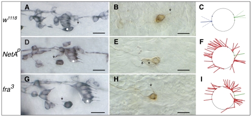
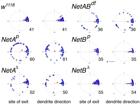
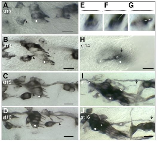
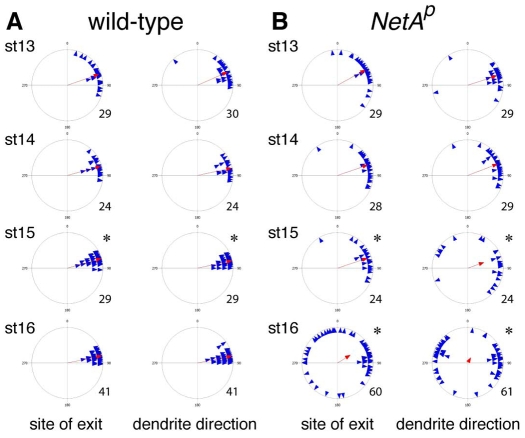
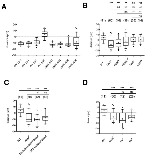
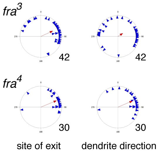
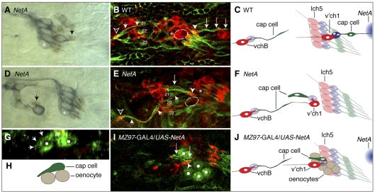
Accessory cells, which include glia and other cell types that develop in close association with neurons, have been shown to play key roles in regulating neuron development. However, the underlying molecular and cellular mechanisms remain poorly understood. A particularly intimate association between accessory cells and neurons is found in insect chordotonal organs. We have found that the cap cell, one of two accessory cells of v'ch1, a chordotonal organ in the Drosophila embryo, strongly influences the development of its associated neuron. As it projects a long dorsally directed cellular extension, the cap cell reorients the dendrite of the v'ch1 neuron and tows its cell body dorsally. Cap cell morphogenesis is regulated by Netrin-A, which is produced by epidermal cells at the destination of the cap cell process. In Netrin-A mutant embryos, the cap cell forms an aberrant, ventrally directed process. As the cap cell maintains a close physical connection with the tip of the dendrite, the latter is dragged into an abnormal position and orientation, and the neuron fails to undergo its normal dorsal migration. Misexpression of Netrin-A in oenocytes, secretory cells that lie ventral to the cap cell, leads to aberrant cap cell morphogenesis, suggesting that Netrin-A acts as an instructive cue to direct the growth of the cap cell process. The netrin receptor Frazzled is required for normal cap cell morphogenesis, and mutant rescue experiments indicate that it acts in a cell-autonomous fashion.


Figures

Fig. 1.
Fig. 1.
Netrin-A and frazzled mutant Drosophila embryos show defects in v'ch1 dendrite position and orientation. (A-I) Single hemisegments of mAb 22C10-immunostained embryos (A,D,G), DiI-labelled v'ch1 neurons (B,E,H) and diagrams showing site of exit and orientation of the v'ch1 dendrite in wild-type (C), NetAp (F) and fra3 (I) mutants. Arrows in A, D and G show the v'ch1 dendrite, white dots show the lch5-5 neuron, and the asterisk in E shows the v'ch1 scolopale cell. (C) The mean and range of dendrite exit position (green dot and dashes on circle, representing the v'ch1 cell body) and orientation (unbroken and broken green lines, respectively) in wild-type embryos (n=41) are shown. Equivalent positions and orientations for the axon are shown in blue. (F,I) Each red line in these diagrams represents the site of exit and orientation of individual dendrites that lie outside the wild-type range of positions and orientations (green). In this and all subsequent figures anterior is up and dorsal is to the right. Scale bars: 10 μm.
Fig. 2.
Fig. 2.
Patterns of v'ch1 dendrite growth in Netrin mutant Drosophila embryos. Each blue triangle in these scatter plots shows the values for an individual dendrite, and the red arrow shows the mean value for that parameter (r value). n values are indicated to the right of each plot.
Fig. 3.
Fig. 3.
Morphogenesis and migration of the v'ch1 neuron and cap cell in wild-type Drosophila embryos. Single hemisegments of embryos were fixed at the developmental stage indicated. (A-G) mAb22C10-stained embryos showing v'ch1 dendrite morphology (arrow). (H-J) P0163-GAL4; UAS-tau-lacZ embryos stained with anti-β-galactosidase, showing morphology of the v'ch1 neuron (asterisk) and cap cell (open circle). White dots in A-D and H-J show the position of the lch5-5 neuron. The arrow in H-J shows the dorsal edge/tip of the v'ch1 cap cell. (E-G) Cross-sectional views of v'ch1 reconstructed from a z-series of images. The black line shows the position of the epidermis. The white line shows the dendrite direction. Scale bars: 10 μm.
Fig. 4.
Fig. 4.
Refinement of v'ch1 dendrite position and orientation does not occur in Netrin-A Drosophila embryos. Each blue triangle in these scatter plots shows the values for an individual dendrite, and the red arrow shows the mean value for that parameter (r value). n values are indicated to the right of each plot. (A) Asterisks indicate that the distributions at stage 15 are significantly different from previous stages (Watson's U2 test, P<0.05). (B) Asterisks indicate that the distributions at stage 15 and 16 are significantly different from wild-type at equivalent stages (Watson's U2 test, P<0.05).
Fig. 5.
Fig. 5.
Failure of v'ch1 neuron migration following loss of function or mis/overexpression of Netrin-A or frazzled. (A-D) Data in boxplot format (+, mean; middle line, median; box, 25-75% quartiles; error bars, data within 5-95% range; dots, outliers) showing the distance between v'ch1 and lch5-5 neuron cell bodies in: (A) wild-type and NetA mutant embryos at various developmental stages; (B) wild-type and various NetA and NetB mutant embryos at stage 16; (C) wild-type, NetA mutant and NetA misexpression embryos at stage 16; (D) wild-type, NetAp, fra3 and fra4 mutant embryos at stage 16. A negative value indicates that v'ch1 lies ventral to lch5-5, a positive value that it lies dorsal. Annotations above the charts in B-D indicate whether the value for the genotype beneath is significantly different to that of the genotype on the far left of the accompanying line (***, P<0.001; **, P<0.01; ns, not significant; one-way ANOVA followed by Tukey's post-test comparison). n values for each genotype are shown in brackets.
Fig. 6.
Fig. 6.
Patterns of v'ch1 dendrite growth in frazzled mutants. Each blue triangle in these scatter plots shows the values for an individual dendrite, and the red arrow shows the mean value for that parameter (r value). n values are indicated to the right of each plot.
Fig. 7.
Fig. 7.
Aberrant morphogenesis of v'ch1 cap cell following loss of function or misexpression of Netrin-A. Images of stage-16 Drosophila embryos with the genotypes indicated. (A,D) The scolopale cell nucleus (arrow) is revealed by anti-Prospero immunohistochemistry (brown), and sensory neurons are labelled with mAb 22C10 (blue). The scolopale cell associates closely with the dendrite, whether it is in a normal (A) or aberrant (D) position. (B,E) Cap cells are stained with anti-β-tubulin (green) and neurons with anti-HRP (red). The v'ch1 neuron in the NetA embryo in E (outlined, not included in projection for sake of clarity) has stalled ventral to the lch5 cluster (asterisk); compare with its normal position in the wild-type embryo (B). The cap cell (large arrows) retains its association with the v'ch1 dendrite (arrowhead) in the mutant, but it is oriented anteroventrally and attaches to the epidermis near the cap cell of vchB (small arrows), close to the muscle 21 and 22 attachment sites. The vchB dendrite in B and E is indicated by an open arrowhead. (G,I) NetA was ectopically expressed in oenocytes (white dots) using the MZ97-GAL4 driver line. (G) A transverse view reveals that the cell body of the v'ch1 cap cell (asterisk) has remained in the epidermal layer, directly superficial to the oenocytes and has extended an aberrant ventral process (arrows) along the surface of an oenocyte. (I) The v'ch1 cap cell has projected an aberrant ventral process (arrows) but the nucleus has not translocated into this process. (C,F,H,J) Schematics of B,E,G,I are shown in C,F,H,J, respectively. The v'ch1 neuron cell body is outlined. The site of endogenous NetA expression is shown in blue in C and J. Scale bars: 10 μm.

References

    1. Bhat K. M., Gaziova I., Krishnan S. (2007). Regulation of axon guidance by slit and netrin signaling in the Drosophila ventral nerve cord. Genetics 176, 2235-2246 - PMC - PubMed
    1. Bradford D., Cole S. J., Cooper H. M. (2009). Netrin-1: diversity in development. Int. J. Biochem. Cell Biol. 41, 487-493 - PubMed
    1. Campos-Ortega J. A., Hartenstein V. (1985). The embryonic development of Drosophila melanogaster Berlin: Springer Verlag;
    1. Carlson S. D., Hilgers S. L., Juang J. L. (1997a). First developmental signs of the scolopale (glial) cell and neuron comprising the chordotonal organ in the Drosophila embryo. Glia 19, 269-274 - PubMed
    1. Carlson S. D., Hilgers S. L., Juang J. L. (1997b). Ultrastructure and blood-nerve barrier of chordotonal organs in the Drosophila embryo. J. Neurocytol. 26, 377-388 - PubMed

Publication types