

Visualizing extravasation dynamics of metastatic tumor cells
- PMID: 20530574
- PMCID: PMC2886748
- DOI: 10.1242/jcs.069443
Visualizing extravasation dynamics of metastatic tumor cells
Abstract
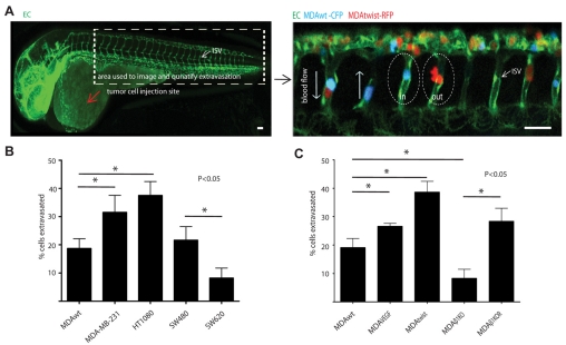
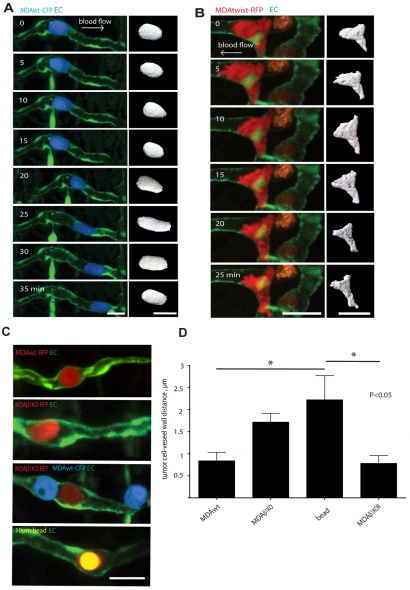
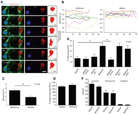
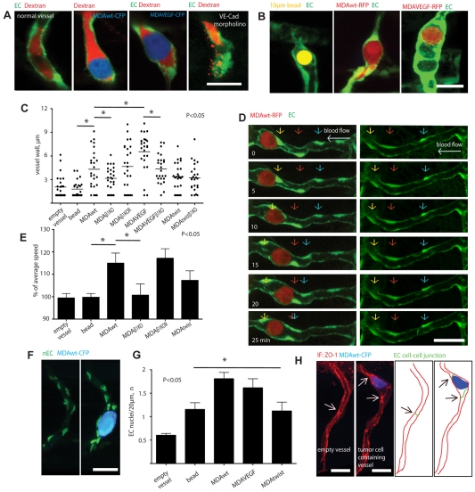
Little is known about how metastatic cancer cells arrest in small capillaries and traverse the vascular wall during extravasation in vivo. Using real-time intravital imaging of human tumor cells transplanted into transparent zebrafish, we show here that extravasation of cancer cells is a highly dynamic process that involves the modulation of tumor cell adhesion to the endothelium and intravascular cell migration along the luminal surface of the vascular wall. Tumor cells do not damage or induce vascular leak at the site of extravasation, but rather induce local vessel remodeling characterized by clustering of endothelial cells and cell-cell junctions. Intravascular locomotion of tumor cells is independent of the direction of blood flow and requires beta1-integrin-mediated adhesion to the blood-vessel wall. Interestingly, the expression of the pro-metastatic gene Twist in tumor cells increases their intravascular migration and extravasation through the vessel wall. However, in this case, Twist expression causes the tumor cells to switch to a beta1-integrin-independent mode of extravasation that is associated with the formation of large dynamic rounded membrane protrusions. Our results demonstrate that extravasation of tumor cells is a highly dynamic process influenced by metastatic genes that target adhesion and intravascular migration of tumor cells, and induce endothelial remodeling.
Figures
Similar articles
-
The Sal-like 4 - integrin α6β1 network promotes cell migration for metastasis via activation of focal adhesion dynamics in basal-like breast cancer cells.Biochim Biophys Acta Mol Cell Res. 2017 Jan;1864(1):76-88. doi: 10.1016/j.bbamcr.2016.10.012. Epub 2016 Oct 20. Biochim Biophys Acta Mol Cell Res. 2017. PMID: 27773610
-
High-resolution imaging of the dynamic tumor cell vascular interface in transparent zebrafish.Proc Natl Acad Sci U S A. 2007 Oct 30;104(44):17406-11. doi: 10.1073/pnas.0703446104. Epub 2007 Oct 22. Proc Natl Acad Sci U S A. 2007. PMID: 17954920 Free PMC article.
-
Impairing flow-mediated endothelial remodeling reduces extravasation of tumor cells.Sci Rep. 2021 Jun 23;11(1):13144. doi: 10.1038/s41598-021-92515-2. Sci Rep. 2021. PMID: 34162963 Free PMC article.
-
Extravasation of immune and tumor cells from an endothelial perspective.J Cell Sci. 2024 Nov 1;137(21):jcs262066. doi: 10.1242/jcs.262066. Epub 2024 Nov 12. J Cell Sci. 2024. PMID: 39530179 Review.
-
The contribution of platelets to intravascular arrest, extravasation, and outgrowth of disseminated tumor cells.Clin Exp Metastasis. 2020 Feb;37(1):47-67. doi: 10.1007/s10585-019-10009-y. Epub 2019 Nov 22. Clin Exp Metastasis. 2020. PMID: 31758288 Review.
Cited by
-
YAP localization mediates mechanical adaptation of human cancer cells during extravasation in vivo.bioRxiv [Preprint]. 2023 Nov 16:2023.11.14.567015. doi: 10.1101/2023.11.14.567015. bioRxiv. 2023. PMID: 38076880 Free PMC article. Preprint.
-
Cancer Cell Invasion and Metastasis in Zebrafish Models (Danio rerio).Methods Mol Biol. 2021;2294:3-16. doi: 10.1007/978-1-0716-1350-4_1. Methods Mol Biol. 2021. PMID: 33742390 Free PMC article.
-
Emergence of zebrafish models in oncology for validating novel anticancer drug targets and nanomaterials.Drug Discov Today. 2013 Feb;18(3-4):128-40. doi: 10.1016/j.drudis.2012.08.002. Epub 2012 Aug 10. Drug Discov Today. 2013. PMID: 22903142 Free PMC article. Review.
-
3D functional and perfusable microvascular networks for organotypic microfluidic models.J Mater Sci Mater Med. 2015 May;26(5):180. doi: 10.1007/s10856-015-5520-5. Epub 2015 Apr 17. J Mater Sci Mater Med. 2015. PMID: 25893395 Review.
-
Pilot Study of an Integrative New Tool for Studying Clinical Outcome Discrimination in Acute Leukemia.Front Oncol. 2019 Apr 9;9:245. doi: 10.3389/fonc.2019.00245. eCollection 2019. Front Oncol. 2019. PMID: 31024847 Free PMC article.
References
-
- Al-Mehdi A. B., Tozawa K., Fisher A. B., Shientag L., Lee A., Muschel R. J. (2000). Intravascular origin of metastasis from the proliferation of endothelium-attached tumor cells: a new model for metastasis. Nat. Med. 6, 100-102 - PubMed
-
- Blum Y., Belting H. G., Ellertsdottir E., Herwig L., Lüders F., Affolter M. (2008). Complex cell rearrangements during intersegmental vessel sprouting and vessel fusion in the zebrafish embryo. Dev. Biol. 316, 312-322 - PubMed
-
- Colmone A., Amorim M., Andrea L., Pontier A. L., Wang S., Jablonski E., Sipkins D. A. (2008). Leukemic cells create bone marrow niches that disrupt the behavior of normal hematopoietic progenitor cells. Science 322, 1861-1865 - PubMed
-
- Crawford Y., Ferrara N. (2008). VEGF inhibition: insights from preclinical and clinical studies. Cell Tissue Res. 335, 261-269 - PubMed
Publication types
MeSH terms
Substances
Grants and funding
LinkOut - more resources
Full Text Sources
Other Literature Sources
Molecular Biology Databases
