. 2010 Aug 15;19(16):3206-18.
doi: 10.1093/hmg/ddq230. Epub 2010 Jun 8.

Neurotoxic effects of TDP-43 overexpression in C. elegans

Affiliations

Neurotoxic effects of TDP-43 overexpression in C. elegans

Peter E A Ash et al. Hum Mol Genet. .

Abstract

RNA-binding protein TDP-43 has been associated with multiple neurodegenerative diseases, including amyotrophic lateral sclerosis and frontotemporal lobar dementia. We have engineered pan-neuronal expression of human TDP-43 protein in Caenorhabditis elegans, with the goal of generating a convenient in vivo model of TDP-43 function and neurotoxicity. Transgenic worms with the neuronal expression of human TDP-43 exhibit an 'uncoordinated' phenotype and have abnormal motorneuron synapses. Caenorhabditis elegans contains a single putative ortholog of TDP-43, designated TDP-1, which we show can support alternative splicing of CFTR in a cell-based assay. Neuronal overexpression of TDP-1 also results in an uncoordinated phenotype, while genetic deletion of the tdp-1 gene does not affect movement or alter motorneuron synapses. By using the uncoordinated phenotype as a read-out of TDP-43 overexpression neurotoxicty, we have investigated the contribution of specific TDP-43 domains and subcellular localization to toxicity. Full-length (wild-type) human TDP-43 expressed in C. elegans is localized to the nucleus. Deletion of either RNA recognition domain (RRM1 or RRM2) completely blocks neurotoxicity, as does deletion of the C-terminal region. These deleted TDP-43 variants still accumulate in the nucleus, although their subnuclear distribution is altered. Interestingly, fusion of TDP-1 C-terminal sequences to TDP-43 missing its C-terminal domain restores normal subnuclear localization and toxicity in C. elegans and CFTR splicing in cell-based assays. Overexpression of wild-type, full-length TDP-43 in mammalian cells (differentiated M17 cells) can also result in cell toxicity. Our results demonstrate that in vivo TDP-43 neurotoxicity can result from nuclear activity of overexpressed full-length protein.


Figures

Figure 1.
Figure 1.
Comparison of hTDP-43 and TDP-1 domain structure. Human TDP-43 and C. elegans TDP-1 have similar arrangements of nuclear localization signals, RRMs and candidate caspase-cleavage sites (arrows). However, TDP-1 lacks an apparent glycine-rich domain and has an extended N-terminus.
Figure 2.
Figure 2.
Expression of human TDP-43 in C. elegans. (A) Comparison of plate phenotypes for wild-type and snb-1/hTDP-43 transgenic worms. Note irregular movement tracks and non-sinusoidal body posture of the snb-1/hTDP-43 worm which is never observed in wild-type animals. (B) Quantification of movement defects in adult snb-1/hTDP-43 worms (strain CL2609), assayed by measuring total distance traveled for individual worms over 40 s interval. (C) Anterior portion of fixed and permeabilized snb-1/hTDP-43 worm probed with anti-hTDP-43 antibody (ProteinTech anti-TARDBP polyclonal antibody). Note specific localization of TDP-43 to the nuclei of nerve ring neurons. Red, anti-hTDP-43; blue, DAPI staining of nuclei; green, intestinal GFP from transformation marker plasmid. (D) Immunoblot of extracts from wild-type and snb-1/hTDP-43 transgenic worms (strain CL1682) probed with anti-TDP-43 monoclonal antibody M01. Note single anti-TDP-43-reactive band in snb-1/hTDP-43 worms.
Figure 3.
Figure 3.
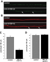
Neuropathology in snb-1/hTDP-43 transgenic worms. (A) Visualization of GABAnergic motor neuron synapses in dorsal cord of living control (CL1685) and snb-1/hTDP-43 (CL1681) worms using an unc-25/SNB-1::GFP reporter transgene (juIs1). Synapses are visualized as concentrations of the reporter along the axons (‘beads on a string’); note significant reductions in detectable synapses in snb-1/hTDP-43 worm (arrows). (B) Dorsal cord axonal processes in wild-type and snb-1/hTDP-43 worms, visualized by co-injection with rgef-1/DsRed2, a reporter that accumulates in all axonal processes (40). Note defasiculations in snb-1/hTDP-43 axonal bundles (arrows). (C) Quantification of dorsal GABAnergic synapses in control transgenic (CL1685) and snb-1/hTDP-43 (CL1681) worms, 30 animals scored for each strain. CL1685, 15.7 ± 0.7 puncta/100 µm; CL1681, 6.5 ± 0.7 puncta/100 µm, Student's t-test P < 0.0001. (D) Quantification of dorsal GABAnergic synapses in control (juIs1) and tdp-1(ok781); juIs1 worms, 30 animals scored for each strain. juIs1, 18.4 ± 0.4 puncta/100 µm; tdp-1(ok781); juIs1, 18.7 ± 0.4 puncta/100 µm, Student's t-test P > 0.5. Error bars = SEM.
Figure 4.
Figure 4.
The uncoordinated phenotype induced by snb-1/hTDP-43 is not associated with neuronal loss. (A) Visualization of GABAnergic motor neurons using unc-47/DsRed2 reporter transgene (hdIs22). (B) Quantification of GABAnergic motor neurons in control and snb-1/hTDP-43 worms, 30 animals scored for each strain. hdIs22 control, 17.6 ± 0.2 GABAnergic motorneurons per worm; hdIs22; snb-1/hTDP-43, 17.7 ± 0.2 GABAnergic motorneurons per worm, Student's t-test P > 0.5. (C) Effect of ced-4(n1162) and tdp-1(ok781) on movement in liquid in snb-1/hTDP-43 worms. ced-4(n1162) does not influence the movement in non-transgenic [wild-type, 47.8 ± 0.6 thrashes/30 s; ced-4(n1162), 48.7 ± 1.8 thrashes/30 s, Student's t-test P > 0.5] or transgenic [snb-1/hTDP-43, 9.4 ± 0.8 thrashes/30 s; ced-4(n1162); snb-1/hTDP-43, 7.9 ± 1.1 thrashes/30 s, Student's t-test P > 0.2] worms. Similarly, introduction of the tdp-1(ok781) deletion into the snb-1/hTDP-43 background does not alter the reduced movement caused by this transgene [snb-1/hTDP-43, 9.4 ± 0.8 thrashes/30 s; tdp-1(ok781); snb-1/hTDP-43, 8.3 ± 0.5 thrashes/30 s, Student's t-test P > 0.1]. Error bars = SEM.
Figure 5.
Figure 5.
TDP-1 can promote CFTR alternative splicing in a cell-based assay. (A) Schematic diagram of the CFTR C155T minigene transfected in add-back assay (dotted lines represent possible splicing outcomes). (B) Effect on CFTR exon 9 splicing of adding back siRNA-resistant wild-type human TDP-43 and TDP-1 following knockdown of the endogenous TDP-43. Standard deviations obtained in three independent transfection experiments are shown. The western blots against the endogenous TDP-43 and tubulin are shown in the lower boxes to show silencing efficiency and equal loading. The weak immunoblot signal from the transfected siRNA-resistant TDP-43 is likely due to the relatively low transfection efficiency of this plasmid construct (37).
Figure 6.
Figure 6.
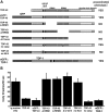
Neurotoxicity of engineered TDP-43 variants expressed in C. elegans. (A) Schematic representation of TDP-43 variants expressed in C. elegans. (B) Quantification of movement defects induced by the expression of wild-type or variant TDP-43. Transgenic worms expressing wild-type TDP-43 (strain CL1682), eGFP::TDP-43 (strain CL1626), TDP-43 caspase site mutant (strain CL1690) or eGFP::TDP-1 (strain LEN144) all show significantly reduced body thrashes in liquid in comparison to control transgenic worms expressing a marker transgene only (strain CL1685) (P < 0.001, Student's t-test). In contrast, transgenic worms expressing eGFP::TDP-43 with a deletion of RRM1 (strain CL1702), RRM2 (strain CL1705) or the C-terminus (strain CL1710) do not have reduced body thrash rates (t-test P > 0.2). Mutation of the nuclear localization signal in an eGFP::TDP-43 construct (strain CL1687) similarly restores body movement to control levels. All strains were assayed at 16°C, except LEN144, which was assayed at 25°C to maximize transgene expression. Error bars = SEM.
Figure 7.
Figure 7.
Localization of snb-1-driven full-length and variant hTDP-43 constructs expressed in C. elegans. (AF) Fused GFP/DIC images of live transgenic worms expressing eGFP::TDP-43 fusions (main panel), with complementary inserts of the nerve ring area of a fixed worm from the same transgenic strain probed with anti-TDP-43 antibody (red) and counter-stained with DAPI (blue) to highlight nuclei. (A) Nerve ring area of eGFP::hTDP-43 transgenic worm (CL1626). GFP fluorescence and anti-TDP-43 staining were localized to nuclei and were primarily diffuse in the nucleus. (B) Nerve ring area of eGFP::hTDP-43 RRM1 deletion transgenic worm (CL1702). GFP fluorescence and anti-TDP-43 staining were nuclear but more punctate than observed for full-length eGFP::hTDP-43. (C) Nerve ring area of eGFP::hTDP-43 RRM2 deletion transgenic worm (CL1705). GFP fluorescence and anti-TDP-43 staining were nuclear but also somewhat more punctate than observed for full-length eGFP::hTDP-43. (D) Nerve ring area of eGFP::hTDP-43 C-terminal deletion (i.e. hTDP-43 1–257, strain CL1710). GFP fluorescence and anti-TDP-43 staining were localized to a single nuclear body/inclusion in most neurons. (E) Nerve ring area of eGFP::hTDP-43 no caspase-cleavage (D89E, D219E) mutant transgenic worm (CL1690). As observed for the eGFP::hTDP-43 (wild-type) fusion (A), GFP fluorescence and anti-TDP-43 staining were localized to nuclei and were primarily diffuse in the nucleus. (F) Nerve ring area of eGFP::hTDP-43 NLS1 mutant transgenic worm (strain CL1687). GFP fluorescence and anti-TDP-43 staining were observed in both perinuclear cytoplasmic inclusions and diffusely in the cytoplasm. (G) Nerve ring area of fixed worm expressing an hTDP-43::TDP-1 fusion construct (CL1714), probed with anti-hTDP-43 monoclonal antibody (red) and DAPI (blue). Addition of C-terminal TDP-1 sequences restored diffuse nuclear distribution to this hTDP-43 variant, which is normally lost when hTDP-43 C-terminal sequences are deleted (compare with D inset). (H) Ventral cord region of fixed eGFP::hTDP-43 transgenic worm (CL1626) (GFP in green, DAPI staining, false-colored red). The fusion protein shows strict nuclear localization, as illustrated by comparison of GFP + DAPI (left) and DAPI only (right) images (arrows). (I) Ventral cord region of fixed eGFP::hTDP-25 F1 transgenic worm (GFP in green, DAPI staining false, colored red). The fusion protein forms large inclusions with strict cytoplasmic localization, as illustrated by comparison of GFP + DAPI (left) and DAPI only (right) images (arrows). Size bar = 10 µm in all images.
Figure 8.
Figure 8.
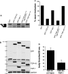
Activity of TDP-43 deletion and fusion proteins. (A) cDNA prepared from co-transfected cells was used as a template for PCR to determine relative exclusion of CFTR exon 9 from the (TG)13(T)5 minigene. (B) Immunoblot using (rabbit) anti-GFP (Invitrogen) to probe for tagged construct expression. Loading controlled for using (mouse) anti-GAPDH (BioDesign). (C) Desitometric readings from CFTR minigene PCR (A) were used to determine the percent inclusion of exon 9 in CFTR minigene for each sample. [In contrast to the pTB CFTR C155T minigene used in Figure 5A, the (TG)13(T)5 CFTR construct used to assess the function of the TDP-43aa1–270::TDP-1aa347–411 fusion construct lacks the C155T CFTR mutation. Under control of endogenous TDP-43 the (TG)13(T)5 minigene is more resistant to inhibition of exon 9 inclusion and requires overexpression of TDP-43 to induce exon 9 skipping (13), providing a rigourous test of the ability of the TDP-43::TDP-1 fusion to promote exon skipping.] (D) Effect of TDP-43::TDP-1 fusion protein on impaired body movement in C. elegans. Transgenic worms containing an addition of TDP-1 C-terminal sequences to the TDP-43 N-terminus have a significant decrease in body movement (P < 0.01, Student's t-test).
Figure 9.
Figure 9.
Toxicity of full-length hTDP-43 in mammalian cell culture. Differentiated M17 cells were transfected for 72 h with 1 µg of constructs expressing either wild-type or tagged full-length hTDP-43. (A) Visualization of cellular distributions of transfected proteins. Top left panel, GFP fluorescence; top right panel, anti-TDP-43 (ProteinTech polyclonal antibody) immunofluorescence; bottom left panel, anti-myc immunofluorescence; bottom right panel, anti-FLAG immunofluorescence. All detected transfected protein expression was nuclear. Size bar = 10 µm. (B) TDP-43 expression levels of transfected cells measured by immunoblot (polyclonal anti-TDP-43 antibody, ProteinTech). (C) Densitometric analysis of immunoblot shown in (B). Note transfected cells express ∼2–3-fold more TDP-43 than control transfected cells. (D) Cell toxicity of TDP-43 overexpression assayed by LDH release. LDH release was significantly higher in cells transfected with TDP-43 constructs than in control vector-transfected cells. Data from three separate experiments were analyzed by one-way analysis of variance followed by Tukey's post hoc analysis. (***P < 0.001).

References

    1. Ou S.H., Wu F., Harrich D., Garcia-Martinez L.F., Gaynor R.B. Cloning and characterization of a novel cellular protein, TDP-43, that binds to human immunodeficiency virus type 1 TAR DNA sequence motifs. J. Virol. 1995;69:3584–3596. - PMC - PubMed
    1. Buratti E., Baralle F.E. Characterization and functional implications of the RNA binding properties of nuclear factor TDP-43, a novel splicing regulator of CFTR exon 9. J. Biol. Chem. 2001;276:36337–36343. doi:10.1074/jbc.M104236200. - DOI - PubMed
    1. Strong M.J., Volkening K., Hammond R., Yang W., Strong W., Leystra-Lantz C., Shoesmith C. TDP43 is a human low molecular weight neurofilament (hNFL) mRNA-binding protein. Mol. Cell Neurosci. 2007;35:320–327. doi:10.1016/j.mcn.2007.03.007. - DOI - PubMed
    1. Arai T., Hasegawa M., Akiyama H., Ikeda K., Nonaka T., Mori H., Mann D., Tsuchiya K., Yoshida M., Hashizume Y., Oda T. TDP-43 is a component of ubiquitin-positive tau-negative inclusions in frontotemporal lobar degeneration and amyotrophic lateral sclerosis. Biochem. Biophys. Res. Commun. 2006;351:602–611. doi:10.1016/j.bbrc.2006.10.093. - DOI - PubMed
    1. Neumann M., Sampathu D.M., Kwong L.K., Truax A.C., Micsenyi M.C., Chou T.T., Bruce J., Schuck T., Grossman M., Clark C.M., et al. Ubiquitinated TDP-43 in frontotemporal lobar degeneration and amyotrophic lateral sclerosis. Science. 2006;314:130–133. doi:10.1126/science.1134108. - DOI - PubMed

Publication types

MeSH terms

Substances