. 2010 Jun 15;70(12):5064-73.
doi: 10.1158/0008-5472.CAN-09-3307. Epub 2010 Jun 8.

E6-associated protein is required for human papillomavirus type 16 E6 to cause cervical cancer in mice

Affiliations

E6-associated protein is required for human papillomavirus type 16 E6 to cause cervical cancer in mice

Anny Shai et al. Cancer Res. .

Abstract

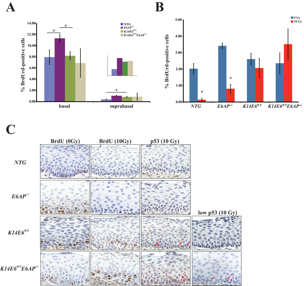
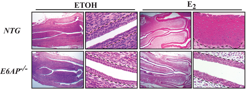
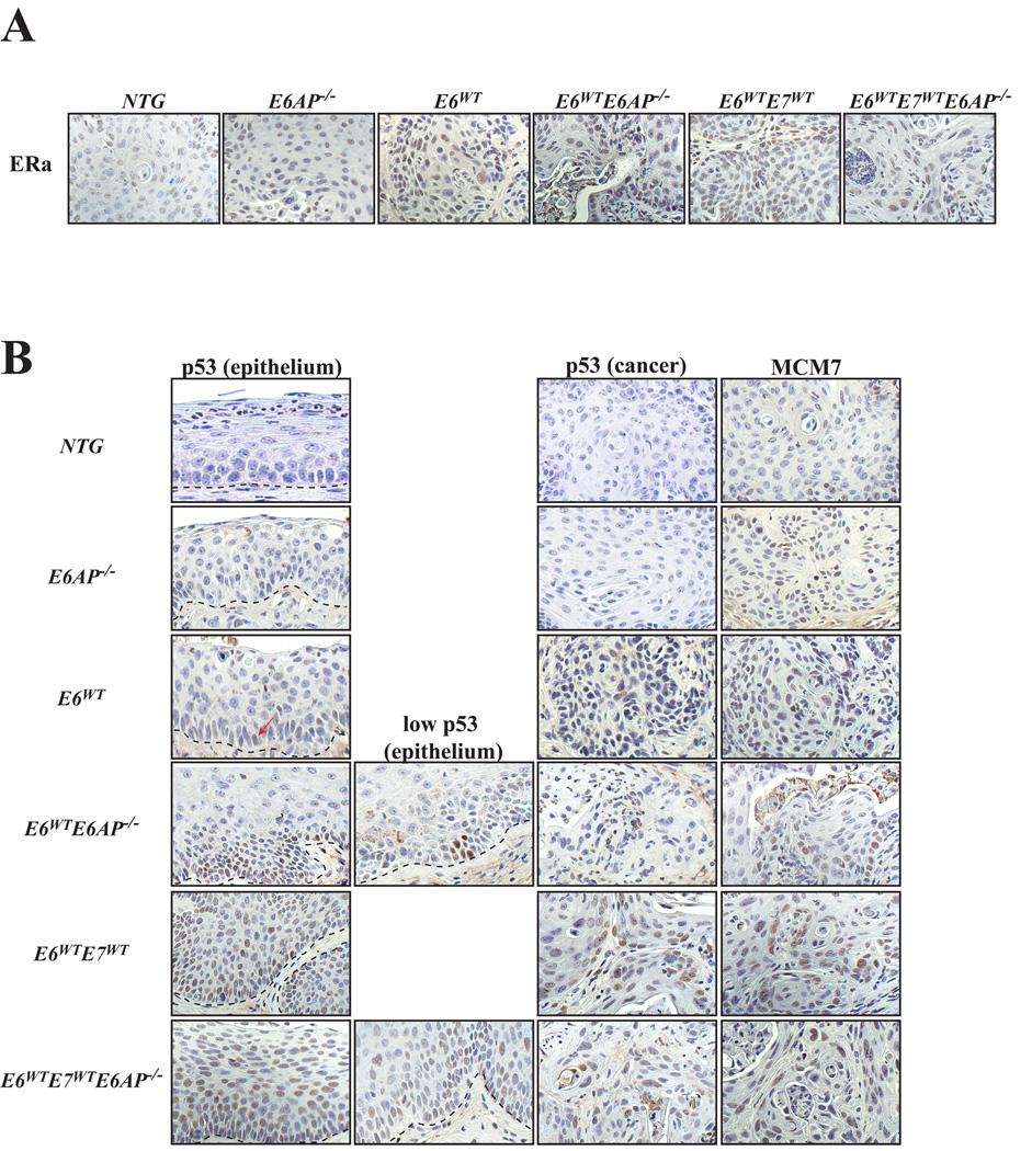
High-risk human papillomaviruses (HPV) cause certain anogenital and head and neck cancers. E6, one of three potent HPV oncogenes that contribute to the development of these malignancies, is a multifunctional protein with many biochemical activities. Among these activities are its ability to bind and inactivate the cellular tumor suppressor p53, induce expression of telomerase, and bind to various other proteins, including Bak, E6BP1, and E6TP1, and proteins that contain PDZ domains, such as hScrib and hDlg. Many of these activities are thought to contribute to the role of E6 in carcinogenesis. The interaction of E6 with many of these cellular proteins, including p53, leads to their destabilization. This property is mediated at least in part through the ability of E6 to recruit the ubiquitin ligase E6-associated protein (E6AP) into complexes with these cellular proteins, resulting in their ubiquitin-mediated degradation by the proteasome. In this study, we address the requirement for E6AP in mediating acute and oncogenic phenotypes of E6, including induction of epithelial hyperplasia, abrogation of DNA damage response, and induction of cervical cancer. Loss of E6AP had no discernible effect on the ability of E6 to induce hyperplasia or abrogate DNA damage responses, akin to what we had earlier observed in the mouse epidermis. Nevertheless, in cervical carcinogenesis studies, there was a complete loss of the oncogenic potential of E6 in mice nulligenic for E6AP. Thus, E6AP is absolutely required for E6 to cause cervical cancer.


Figures

Figure 1
Figure 1. Examination of acute effects in HPV E6 transgenic mice on an E6AP-deficient background in the cervix
A. The graph shows the quantification of DNA synthesis in the cervical epithelium of female mice acutely treated with estrogen to synchronize mice in estrus. Groups of three or more mice per genotype were injected with BrdUrd one hour prior to sacrifice. Formalin fixed, paraffin embedded (FFPE) tissue was subjected to BrdUrd-specific immunohistochemistry and the percentage of BrdUrd-positive cells were counted both in the basal and suprabasal layers in at least eight 40× frames/mouse tissue, as a measure of DNA synthesis. Epithelial hyperplasia is defined by an increase in the percentage of suprabasal BrdUrd-positive cells. E6AP−/− mice have a marginal increase in the number of basal BrdUrd-positive cells relative to NTG (p=0.08, #) mice and is significantly different than K14E6WT mice (p=0.03, *). The percentage of suprabasal BrdUrd-positive cells was significantly higher in both E6AP−/− and K14E6WT groups (p<0.05) relative to NTG mice. Loss of E6AP in the presence of HPV E6 did not prevent the induction of epithelial hyperplasia (p=1). The inset is a magnification of the number of the BrdUrd-positive cells in the suprabasal layers. B. The graph shows the quantification of DNA synthesis in the cervical epithelium of females acutely treated with estrogen and either given 10Gy of ionizing radiation or not treated. Groups of three or more mice per genotype were injected with BrdUrd 23 hours post irradiation and one hour later tissue was harvested. Like size groups of age matched mice not exposed to irradiation served as controls. Percentage BrdUrd-positive cells was quantified as described in A. Following irradiation, significant reductions in the percentage of cells supporting DNA synthesis were observed in both NTG (*, p=0.01) and E6AP−/− (*, p=0.03) groups. Both K14E6 WT and K14E6 WTE6AP−/− groups were abrogated in this DNA damage response, maintaining the levels of DNA synthesis seen in the control groups for each genotype (both p>0.05). C. Shown are sections of cervical epithelium from mice acutely treated with exogenous estrogen and stained for BrdUrd or p53 by IHC. Mice were either unirradiated or irradiated and sacrificed 24hours later. Dotted lines in each picture demarcate the cervical epithelium from the underlying stroma. DNA synthesis, as represented by BrdUrd positive staining nuclei, is reduced upon radiation in nontransgenic mice on either an E6AP-sufficient or insufficient background (first and second columns). In contrast, both K14E6WT and K14E6WTE6AP−/− retain the ability to synthesize DNA. p53 is undetectable in the absence of DNA damage (data not shown), however is induced in NTG and E6AP−/− mice upon radiation (third column). K14E6WT and K14E6WTE6AP−/− mice display variable levels of p53 upon irradiation. Arrows point to examples of p53-positive cells. Examples of a strong inhibition of the DNA damage response as exemplified by either a lack of (K14E6WT) or a few (K14E6WTE6AP−/−) p53-positive cells in the epithelium are also shown (fourth column).
Figure 2
Figure 2. Acute responses to estrogen partially require E6AP
Shown are H/E sections of cervix from mice that were ovariectomized and treated either with ETOH or estrogen (E2) for 24hours. The first and third columns are sections taken at 1.25× magnification, while the second and fourth columns are sections taken at 40× magnification. Note the relative lack of epithelial hyperplasia in E6AP−/− mice relative to NTG mice upon treatment with estrogen (far right column).
Figure 3
Figure 3. Immunohistochemical characterization of tumors from the reproductive tract
A. Shown are sections of cancers taken from the reproductive tract from mice of the indicated genotypes and stained for ERα by IHC. All of the cancers from the various genotypes express ERα. All of the images are 40× magnification. B. Shown are sections of epithelium (first & second column) or cancers (third column) taken from the reproductive tract from mice of the indicated genotypes and stained by IHC for either p53 or MCM7. Dotted lines in each picture demarcate the cervical epithelium from the underlying stroma. In the epithelium, p53 is not often detected, however, loss of E6AP in K14E6WTE6AP−/− mice results in the induction of p53 at either low or higher (second column) levels. p53 is detected both in the epithelium and cancers of K14E6WTE7WT and K14E6WTE7WTE6AP−/− as p53 is stabilized by HPV E7. MCM7 (fourth column) is expressed at low levels in cancers from NTG and E6AP−/− mice. Levels of MCM7 are increased in cancers that express HPV oncogenes, E6 and/or E7. All of the images are 40× magnification.

References

    1. Walboomers JM, Jacobs MV, Manos MM, et al. Human papillomavirus is a necessary cause of invasive cervical cancer worldwide. J Pathol. 1999;189:12–19. - PubMed
    1. zur Hausen H. Papillomavirus infections--a major cause of human cancers. Biochim Biophys Acta. 1996;1288:F55–F78. - PubMed
    1. Parkin DM. The global health burden of infection-associated cancers in the year 2002. Int J Cancer. 2006;118:3030–3044. - PubMed
    1. Parkin DM, Bray F. Chapter 2: The burden of HPV-related cancers. Vaccine. 2006;24 Suppl 3 S3/11–25. - PubMed
    1. Sankaranarayanan R, Ferlay J. Worldwide burden of gynaecological cancer: the size of the problem. Best Pract Res Clin Obstet Gynaecol. 2006;20:207–225. - PubMed

Publication types

MeSH terms