Review
. 2010 Jun 15;16(12):3141-52.
doi: 10.1158/1078-0432.CCR-09-2823. Epub 2010 Jun 8.

Targeting Notch to target cancer stem cells

Affiliations
Review

Targeting Notch to target cancer stem cells

Antonio Pannuti et al. Clin Cancer Res. .

Abstract

The cellular heterogeneity of neoplasms has been at the center of considerable interest since the "cancer stem cell hypothesis", originally formulated for hematologic malignancies, was extended to solid tumors. The origins of cancer "stem" cells (CSC) or tumor-initiating cells (TIC; henceforth referred to as CSCs) and the methods to identify them are hotly debated topics. Nevertheless, the existence of subpopulations of tumor cells with stem-like characteristics has significant therapeutic implications. The stem-like phenotype includes indefinite self-replication, pluripotency, and, importantly, resistance to chemotherapeutics. Thus, it is plausible that CSCs, regardless of their origin, may escape standard therapies and cause disease recurrences and/or metastasis after apparently complete remissions. Consequently, the idea of selectively targeting CSCs with novel therapeutics is gaining considerable interest. The Notch pathway is one of the most intensively studied putative therapeutic targets in CSC, and several investigational Notch inhibitors are being developed. However, successful targeting of Notch signaling in CSC will require a thorough understanding of Notch regulation and the context-dependent interactions between Notch and other therapeutically relevant pathways. Understanding these interactions will increase our ability to design rational combination regimens that are more likely to prove safe and effective. Additionally, to determine which patients are most likely to benefit from treatment with Notch-targeting therapeutics, reliable biomarkers to measure pathway activity in CSC from specific tumors will have to be identified and validated. This article summarizes the most recent developments in the field of Notch-targeted cancer therapeutics, with emphasis on CSC.


Figures

Figure 1
Figure 1
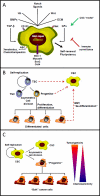
A: schematic representation of an idealized CSC. The figure shows a list, not meant to be all-inclusive, of pathways that modulate the CSC phenotype. CSC exist in the context of “niches” formed by neighboring cells and extracellular matrix ECM). The Hedgehog (Hh), Notch, and Wnt pathways mediate short-range interactions with neighboring cells. Soluble mediators such as TGF-β and the related BMPs, or growth factors such as hepatocyte growth factor (Met ligand), as well as signals from ECM proteins may all participate in regulating the maintenance, self-renewal and differentiation of CSCs. These are characterized by slow replication, ability to generate partially differentiated progenies (pluripotency), highly effective DNA repair, ability to eliminate xenobiotics including chemotherapeutics through ABC family transporters (ABC) and expression of primitive membrane markers (CD133, Met). Transcription factors such as Bmi-1, Musashi, Sox2, Oct4 and others are commonly expressed in putative CSCs. Immune surveillance by the innate and possibly adaptive immune systems also contributes to the CSC microenvironment, with effects that at least in mice are inhibitory. B: Models of CSC origins. In the traditional model, CSC originate from the transformation (red dashed arrows) of normal tissue stem cells (TSC), or possibly of progenitor cells with limited self-replication ability that normally generate cells destined for differentiation. In the alternative model proposed by Mani and colleagues, transformation of cells at many stages of the differentiation process can produce CSC through EMT, which restores a stem-like phenotype -and the ability to metastasize- to some cancer cells. Besides EMT, other mechanisms of de-differentiation have been described and may contribute to restoration of stemness in transformed cells. C: Hierarchical organization of cancers. Once CSC are formed, they are thought to generate other tumor cells through a process akin to normal tissue differentiation. In a widely accepted model, asymmetric cell division of CSC produces pluripotent “progenitors”, which in turn generate one or more bulk tumor cell types through proliferation and aberrant differentiation. CSC and “progenitors” are more tumorigenic in xenografts and less chemosensitive than bulk cancer cells.
Figure 2
Figure 2. Schematic structure of Notch receptors and ligands
Left: Notch ligands contain EGF-like repeats and a trans-membrane domain (TMD) with a short cytoplasmic tail of variable length. The N-terminus contains a specialized DSL region that structurally resembles EGF repeats [108], and a DOS region consisting of two atypical EGF-like repeats. Jagged-1 and -2 (mammalian homologs of Drosophila Serrate) have a longer EGF region than other ligands, and also contain a cyteine-rich motif (CR). Delta homologs in mammals include DSL and DOS-containing ligands (Delta-1) and DSL-only ligands (Delta-3 and -4). It is unclear whether these require a co ligand protein containing a DOS but no DSL (DLK1 and DLK2) to activate Notch. Delta-3 does not activate Notch well in cell culture systems, while Delta-4 does. Right: A typical mature, full length Notch receptor (NFL), in this case human Notch-1, contains an NEC featuring multiple EGF-like repeats (36 in Notch-1). Of these, repeats 11 and 12 (purple) represent the primary ligand binding site, with repeats 24-29 (purple) playing an accessory role. NEC is glycosylated by Pofut-1 and the Fringe enzymes, which add fucose (Fuc) and N-acetylglucosamine (Glc-Nac) to it. RUMI enzymes (so far only identified in Drosophila) add glucose to it. NEC ends with three Lin-Notch repeats (LNR), which fold over the heterodimerization domain (HD), masking the ADAM cleavage site (red) in the N-terminus of NTM. Distal to the HD, NTM contains a transmembrane domain (TMD, shown here with membrane phospholipids surrounding it). The γ-secretase cleavage region is at the cytoplasmic end of the TMD. Moving from N- to C-terminus, we find a RAM (RBP-jκ Activation Motif), a nuclear localization sequence (NLS), seven ankyrin repeats (ANK), and a C-terminal region that contains a PEST (Proline-Glutamic-Serine-Threonine rich) sequence which controls receptor turnover by being phosphorylated by CDK8, leading to ubiquitination by SEL10/Fbw7 and degradation. After ligand-induced subunit separation, the ADAM site is exposed and cleaved by ADAM10 or ADAM17, generating a Notch extracellular truncated (NEXT) intermediate, which is still membrane associated. NEXT is cleaved by γ-secretase, generating NIC.
Figure 3
Figure 3. Diagram of Notch activation and canonical signaling mechanisms
A: Ligand activation and functions. In ligand expressing cells, ligands are ubiqutinated (UQ) by E3 ligases Mindbomb and Neuralized, endocytosed and “activated”. “Active” ligands bind Notch receptors, dissociating NEC from NTM. The complex ligand- NEC is trans-endocytosed into the ligand-expressing cell, perhaps providing mechanical energy to separate NEC from NTM. Some ligands expressed in cis can bind Notch on the same cell, causing cis-inhibition. B: ligand-dependent and -independent activation. Ligand-induced NEC separation unmasks the ADAM cleavage site (red), which is cleaved by ADAM10 or ADAM17, producing NEXT and a short peptide which is released. NEXT is cleaved by γ-secretase, at the membrane or during endocytosis, generating NIC. The release of NIC from endosomes (or the selection of cleavage site by γ-secretase) may require endosome acidification (H+) by aquaporin Bib. The stability of NIC is regulated by factors such as Pin-1 prolyl isomerase and NLK kinase. Endocytosis can lead to ligand-independent Notch activation catalyzed by γ-secretase. In the absence of non-visual β-arrestin Kurz, Deltex may lead to Notch endocytosis and activation. C: control of Notch availability and trafficking. The amount of Notch available at the membrane is controlled by many endocytosis-recycling mechanisms. Several E3 ligases (Itch, CBL, Nedd4, the Deltex-Kurz complex) can target Notch for degradation. The ESCRT complex and lgd in Drosophila (and presumably their homologues in mammals) control Notch degradation, and their loss causes accumulation of Notch in endosomes and ligand-independent activation. In actively dividing cells, Numb/ACBD3 asymmetrically partitions to one daughter cell, causing selective Notch degradation in it. D: nuclear events. NIC is transported to the nucleus, where it causes the dissociation of the co-repressor complex including SHARP, SKIP and several other proteins (CoR) from CSL. Notch, CSL and MAML form a tertiary complex [109] which in turn recruites p300 and other coactivators (CoA) to the chromatin and forming the NTC that activates transcription. The NTC can form heterodimers on the chromatin with other NTCs or supramolecular complexes with other transcription factors, modulating the choice of genes regulated by Notch.
Figure 4
Figure 4. Effects of a GSI on human breast cancer secondary mammospheres
Mammospheres were formed ex vivo from a pleural aspirate from a late-stage breast cancer patient. Mammospheres were dissociated and secondary mammospheres were formed and characterized for multilineage differentiation (an indication they contain pluripotent stem-like cells). Secondary mammospheres were treated with vehicle or a panel of GSIs at clinically achievable concentrations (Grudzien et al., submitted for publication). Mammosphere and isolated cells were enumerated at different times. This example shows phase contrast microphotographs of secondary mammospheres from a clinical isolate treated with vehicle (left) or 10 μM GSI MRK003 for 7 days (right). GSI completely and irreversibly blocked the formation of new mammospheres, and only isolated cells were observed in culture. These eventually underwent apoptosis.

References

    1. Al Hajj M, Becker MW, Wicha M, Weissman I, Clarke MF. Therapeutic implications of cancer stem cells. Curr Opin Genet Dev. 2004;14:43–7. - PubMed
    1. Alison MR, Islam S, Lim SM. Number crunching in the cancer stem cell market. Breast Cancer Res. 2009;11:302. - PMC - PubMed
    1. Donnenberg VS, Donnenberg AD. Multiple drug resistance in cancer revisited: the cancer stem cell hypothesis. J Clin Pharmacol. 2005;45:872–7. - PubMed
    1. Haraguchi N, Inoue H, Tanaka F, et al. Cancer stem cells in human gastrointestinal cancers. Hum Cell. 2006;19:24–9. - PubMed
    1. Hemmati HD, Nakano I, Lazareff JA, et al. Cancerous stem cells can arise from pediatric brain tumors. Proc Natl Acad Sci U S A. 2003;100:15178–83. - PMC - PubMed

Publication types