

Müller cell-derived VEGF is essential for diabetes-induced retinal inflammation and vascular leakage
- PMID: 20530741
- PMCID: PMC2927953
- DOI: 10.2337/db09-1420
Müller cell-derived VEGF is essential for diabetes-induced retinal inflammation and vascular leakage
Abstract
Objective: Vascular endothelial growth factor (VEGF-A or VEGF) is a major pathogenic factor and therapeutic target for diabetic retinopathy (DR). Since VEGF has been proposed as a survival factor for retinal neurons, defining the cellular origin of pathogenic VEGF is necessary for the effectiveness and safety of long-term anti-VEGF therapies for DR. To determine the significance of Müller cell-derived VEGF in DR, we disrupted VEGF in Müller cells with an inducible Cre/lox system and examined diabetes-induced retinal inflammation and vascular leakage in these conditional VEGF knockout (KO) mice.
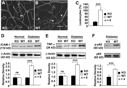
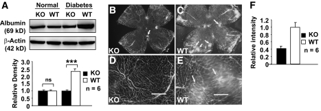
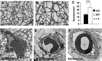
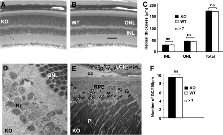
Research design and methods: Leukostasis was determined by counting the number of fluorescently labeled leukocytes inside retinal vasculature. Expression of biomarkers for retinal inflammation was assessed by immunoblotting of TNF-alpha, ICAM-1, and NF-kappaB. Vascular leakage was measured by immunoblotting of retinal albumin and fluorescent microscopic analysis of extravascular albumin. Diabetes-induced vascular alterations were examined by immunoblotting and immunohistochemistry for tight junctions, and by trypsin digestion assays for acellular capillaries. Retinal integrity was analyzed with morphologic and morphometric analyses.
Results: Diabetic conditional VEGF KO mice exhibited significantly reduced leukostasis, expression of inflammatory biomarkers, depletion of tight junction proteins, numbers of acellular capillaries, and vascular leakage compared to diabetic control mice.
Conclusions: Müller cell-derived VEGF plays an essential and causative role in retinal inflammation, vascular lesions, and vascular leakage in DR. Therefore, Müller cells are a primary cellular target for proinflammatory signals that mediates retinal inflammation and vascular leakage in DR.
Figures


Similar articles
-
Ischaemia-induced retinal neovascularisation and diabetic retinopathy in mice with conditional knockout of hypoxia-inducible factor-1 in retinal Müller cells.Diabetologia. 2011 Jun;54(6):1554-66. doi: 10.1007/s00125-011-2081-0. Epub 2011 Mar 1. Diabetologia. 2011. PMID: 21360191 Free PMC article.
-
Müller cell-derived VEGF is a significant contributor to retinal neovascularization.J Pathol. 2009 Dec;219(4):446-54. doi: 10.1002/path.2611. J Pathol. 2009. PMID: 19768732
-
Loss of X-box binding protein 1 in Müller cells augments retinal inflammation in a mouse model of diabetes.Diabetologia. 2019 Mar;62(3):531-543. doi: 10.1007/s00125-018-4776-y. Epub 2019 Jan 6. Diabetologia. 2019. PMID: 30612139 Free PMC article.
-
[Cell biology of intraocular vascular diseases].Nippon Ganka Gakkai Zasshi. 1999 Dec;103(12):923-47. Nippon Ganka Gakkai Zasshi. 1999. PMID: 10643294 Review. Japanese.
-
Functions of Müller cell-derived vascular endothelial growth factor in diabetic retinopathy.World J Diabetes. 2015 Jun 10;6(5):726-33. doi: 10.4239/wjd.v6.i5.726. World J Diabetes. 2015. PMID: 26069721 Free PMC article. Review.
Cited by
-
Retinal Molecular Changes Are Associated with Neuroinflammation and Loss of RGCs in an Experimental Model of Glaucoma.Int J Mol Sci. 2021 Feb 19;22(4):2066. doi: 10.3390/ijms22042066. Int J Mol Sci. 2021. PMID: 33669765 Free PMC article.
-
Oxidative and endoplasmic reticulum stresses mediate apoptosis induced by modified LDL in human retinal Müller cells.Invest Ophthalmol Vis Sci. 2012 Jul 9;53(8):4595-604. doi: 10.1167/iovs.12-9910. Invest Ophthalmol Vis Sci. 2012. PMID: 22678501 Free PMC article.
-
RPE barrier breakdown in diabetic retinopathy: seeing is believing.J Ocul Biol Dis Infor. 2011 Jun;4(1-2):83-92. doi: 10.1007/s12177-011-9068-4. Epub 2011 Dec 31. J Ocul Biol Dis Infor. 2011. PMID: 23275801 Free PMC article.
-
Regulation of inflammation by VEGF/BDNF signaling in mouse retinal Müller glial cells exposed to high glucose.Cell Tissue Res. 2022 Jun;388(3):521-533. doi: 10.1007/s00441-022-03622-z. Epub 2022 Apr 8. Cell Tissue Res. 2022. PMID: 35394215
-
Müller Glial Expression of REDD1 Is Required for Retinal Neurodegeneration and Visual Dysfunction in Diabetic Mice.Diabetes. 2022 May 1;71(5):1051-1062. doi: 10.2337/db21-0853. Diabetes. 2022. PMID: 35167652 Free PMC article.
References
-
- Aiello LP, Avery RL, Arrigg PG, Keyt BA, Jampel HD, Shah ST, Pasquale LR, Thieme H, Iwamoto MA, Park JE, et al. Vascular endothelial growth factor in ocular fluid of patients with diabetic retinopathy and other retinal disorders. N Engl J Med 1994;331:1480–1487 - PubMed
-
- Adamis AP, Miller JW, Bernal MT, D'Amico DJ, Folkman J, Yeo TK, Yeo KT. Increased vascular endothelial growth factor levels in the vitreous of eyes with proliferative diabetic retinopathy. Am J Ophthalmol 1994;118:445–450 - PubMed
-
- Miyamoto K, Khosrof S, Bursell SE, Rohan R, Murata T, Clermont AC, Aiello LP, Ogura Y, Adamis AP. Prevention of leukostasis and vascular leakage in streptozotocin-induced diabetic retinopathy via intercellular adhesion molecule-1 inhibition. Proc Natl Acad Sci U S A 1999;96:10836–10841 - PMC - PubMed
-
- Joussen AM, Poulaki V, Mitsiades N, Kirchhof B, Koizumi K, Dohmen S, Adamis AP. Nonsteroidal anti-inflammatory drugs prevent early diabetic retinopathy via TNF-alpha suppression. FASEB J 2002;16:438–440 - PubMed
Publication types
MeSH terms
Substances
Grants and funding
LinkOut - more resources
Full Text Sources
Medical
Molecular Biology Databases
Research Materials
Miscellaneous
