

Subchondral cystlike lesions develop longitudinally in areas of bone marrow edema-like lesions in patients with or at risk for knee osteoarthritis: detection with MR imaging--the MOST study
- PMID: 20530753
- PMCID: PMC2923728
- DOI: 10.1148/radiol.10091467
Subchondral cystlike lesions develop longitudinally in areas of bone marrow edema-like lesions in patients with or at risk for knee osteoarthritis: detection with MR imaging--the MOST study
Abstract
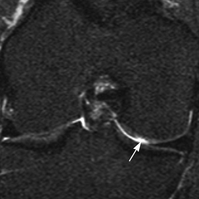
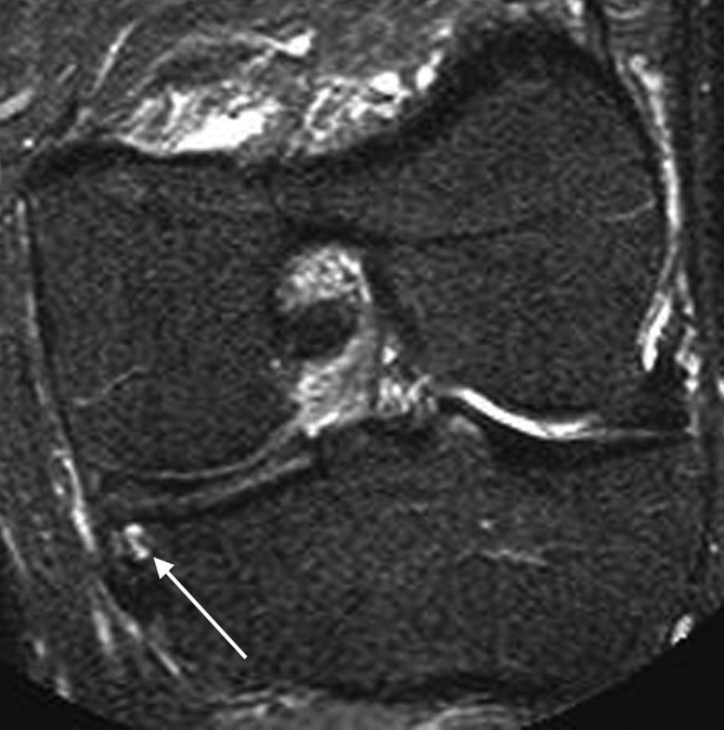
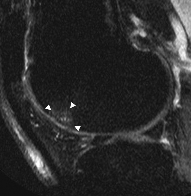
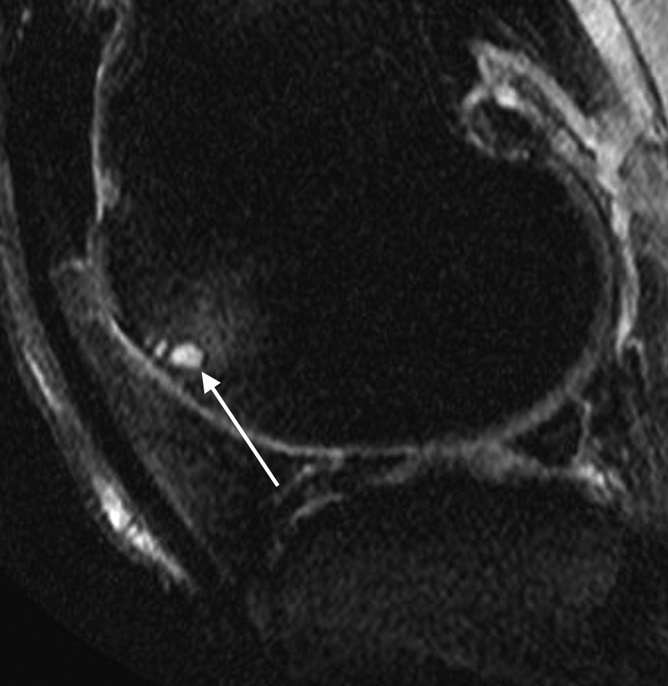
Purpose: To assess the association of prevalent bone marrow edema-like lesions (BMLs) and full-thickness cartilage loss with incident subchondral cyst-like lesions (SCs) in the knee to evaluate the bone contusion versus synovial fluid intrusion theories of SC formation.
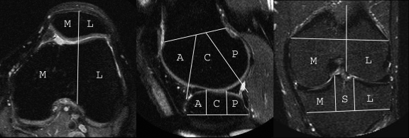
Materials and methods: The Multicenter Osteoarthritis study is a longitudinal study of individuals who have or are at risk for knee osteoarthritis. The HIPAA-compliant protocol was approved by the institutional review boards of all participating centers, and written informed consent was obtained from all participants. Magnetic resonance images were acquired at baseline and 30-month follow-up and read semiquantitatively by using the Whole-Organ Magnetic Resonance Imaging Score system. The tibiofemoral and patellofemoral joints were subdivided into 14 subregions. BMLs and SCs were scored from 0 to 3. Cartilage morphology was scored from 0 to 6. The association of prevalent BMLs and full-thickness cartilage loss with incident SCs in the same subregion was assessed by using logistic regression with mutual adjustment for both predictors.
Results: A total of 1283 knees were included. After adjustment for full-thickness cartilage loss, prevalent BMLs showed a strong and significant association with incident SCs in the same subregion, with an odds ratio of 12.9 (95% confidence interval [CI]: 8.9, 18.6). After adjustment for BMLs, prevalent full-thickness cartilage loss showed a significant but much less important association with incident SCs in the same subregion (odds ratio, 1.4; 95% CI: 1.0, 2.0). There was no apparent relationship between severity of full-thickness cartilage loss at baseline and incident SCs.
Conclusion: Prevalent BMLs strongly predict incident SCs in the same subregion, even after adjustment for full-thickness cartilage loss, which supports the bone contusion theory of SC formation.
(c) RSNA, 2010.
Conflict of interest statement
See Materials and Methods for pertinent disclosures.
Figures

References
-
- Zanetti M, Bruder E, Romero J, Hodler J. Bone marrow edema pattern in osteoarthritic knees: correlation between MR imaging and histologic findings. Radiology 2000;215(3):835–840 - PubMed
-
- Pouders C, De Maeseneer M, Van Roy P, Gielen J, Goossens A, Shahabpour M. Prevalence and MRI-anatomic correlation of bone cysts in osteoarthritic knees. AJR Am J Roentgenol 2008;190(1):17–21 - PubMed
-
- Landells JW. The bone cysts of osteoarthritis. J Bone Joint Surg Br 1953;35-B(4):643–649 - PubMed
-
- Rhaney K, Lamb DW. The cysts of osteoarthritis of the hip: a radiological and pathological study. J Bone Joint Surg Br 1955;37-B(4):663–675 - PubMed
-
- Resnick D, Niwayama G, Coutts RD. Subchondral cysts (geodes) in arthritic disorders: pathologic and radiographic appearance of the hip joint. AJR Am J Roentgenol 1977;128(5):799–806 - PubMed
Publication types
MeSH terms
Grants and funding
LinkOut - more resources
Full Text Sources
Medical
