. 2010 Jun 8;3(125):ra45.
doi: 10.1126/scisignal.2000549.

Purinergic signaling: a fundamental mechanism in neutrophil activation

Affiliations

Purinergic signaling: a fundamental mechanism in neutrophil activation

Yu Chen et al. Sci Signal. .

Abstract

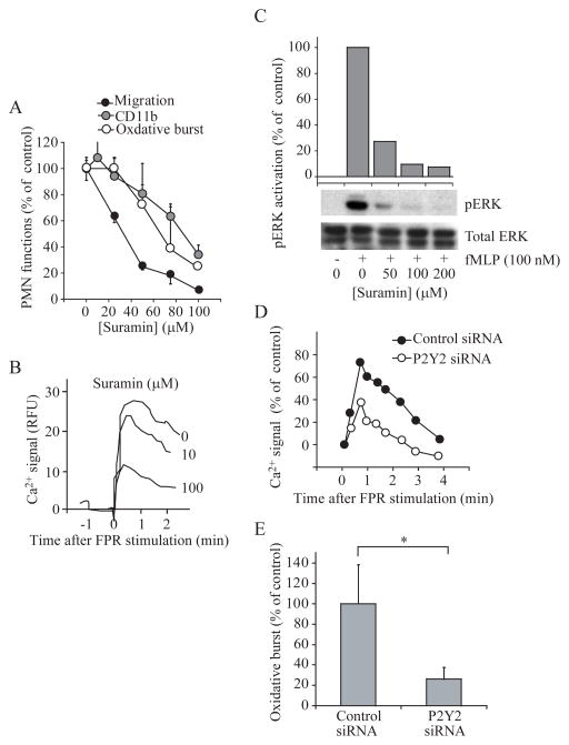
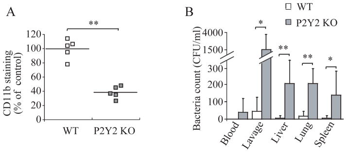
Efficient activation of neutrophils is a key requirement for effective immune responses. We found that neutrophils released cellular adenosine triphosphate (ATP) in response to exogenous stimuli such as formylated bacterial peptides and inflammatory mediators that activated Fcgamma, interleukin-8, C5a complement, and leukotriene B(4) receptors. Stimulation of the formyl peptide receptor (FPR) led to ATP release through pannexin-1 (panx1) hemichannels, and FPRs colocalized with P2Y2 nucleotide receptors on the cell surface to form a purinergic signaling system that facilitated neutrophil activation. Disruption of this purinergic signaling system by inhibiting or silencing panx1 hemichannels or P2Y2 receptors blocked neutrophil activation and impaired innate host responses to bacterial infection. Thus, purinergic signaling is a fundamental mechanism required for neutrophil activation and immune defense.


Conflict of interest statement

Conflicts of interest: The authors have no conflicts of interest to declare.

Figures

Fig. 1
Fig. 1
ATP release channels in PMNs. (A) The abundance of mRNAs for the TTYH1 and TTYH3 maxi-anion channels, connexin 43 (Cx 43), and pannexin-1 (Panx 1) in human brain, primary human PMNs, and HL-60 cells, before and after differentiation (dHL-60) with 1.3% DMSO for 3 days, were estimated by real-time RT-PCR. nt, not tested; nd, not detected. Values are expressed as the mean ± SD, and data are representative of four experiments. (B) Immunostaining of primary human PMNs with antibodies against TTYH1, TTYH3, and Cx 43. (C) Immunostaining of Panx 1 on primary human PMNs that were either unstimulated or were stimulated with fMLP (1 nM). (D) Colocalization of F-actin (red), Panx 1 (green), and FPR (blue) in unpolarized human PMNs and in PMNs that were polarized by stimulation with fMLP (1 nM) that was added uniformly for 10 min. The images shown in panels B and D are representative of experiments performed at least twice with cells from different donors; panel C is representative of at least 3 separate experiments.
Fig. 2
Fig. 2
Maxi-anion and Panx 1 channels mediate the FPR-induced release of ATP from PMNs. (A and B) The effects of DIDS, CBX, 10Panx 1, or a nonsense control peptide on the release of ATP from human PMNs in response to FPR stimulation with 100 nM fMLP for 3 min were examined. (C to E) The effects of DIDS, CBX, 10Panx 1, or nonsense control peptide on the functional responses of human PMNs stimulated with fMLP were determined. (F) Effect of knocking down TTYH3 on ATP release and oxidative burst of dHL-60 cells in response to fMLP. Values in all panels are expressed as the mean ± SD and data are representative of individual experiments with cells from different donors (n ≥ donors), Student’s t-test, *, P < 0.05.
Fig. 3
Fig. 3
FPR-induced Ca2+ mobilization and MAPK activation in human PMNs require the release of ATP. (A) Intracellular Ca2+ mobilization in response to fMLP (10 nM) in human PMNs pretreated with the indicated concentrations of apyrase, CBX or 10Panx 1. Data are expressed as relative fluorescence units (RFU). (B) Effect of pretreatment of human PMNs with the indicated concentrations of CBX or apyrase on fMLP-stimulated activation of ERK, which was determined by assessing the ratio of the abundance of phosphorylated ERK (pERK) to that of total ERK protein. The data are representative of individual experiments with cells from different donors (n = 4 in panel A and 3 in panel B).
Fig. 4
Fig. 4
P2 purinergic receptors are required for FPR-induced cellular responses in human PMNs. (A) The effects of the P2 receptor antagonist suramin on the functional responses of human PMNs to fMLP were examined. (B) The effect of suramin on Ca2+ signaling in PMNs in response to fMLP was examined. (C) The effect of suramin on fMLP-induced activation of ERK in PMNs was determined. (D and E) The effect of knockdown of P2Y2 receptors in dHL-60 cells on FPR-induced Ca2+ signaling and oxidative burst, which was assessed by flow cytometric analysis of Fluo-3 and DHR, respectively, was examined. Data are expressed as a percentage of controls treated with nonsense siRNA. Values are expressed as mean ± SD (A and E) and the data are representative of at least three individual experiments with similar results. *, P < 0.05.
Fig. 5
Fig. 5
FPR and P2Y2 receptors colocalize on the cell surface. (A) dHL-60 cells were cotransfected with plasmids encoding P2Y2-ECFP and FPR-EYFP, and receptor distribution was assessed by confocal laser scanning microscopy. (B) The degree of receptor colocalization in different regions of interest (ROI) was evaluated by regression analysis. ROI 1, ROI 2, and ROI 3 indicate areas with different degrees of receptor colocalization shown in the right-most image in (A). The data are representative of more than three individual experiments.
Fig. 6
Fig. 6
ATP release and autocrine feedback through purinergic receptors are a general requirement for the activation of PMNs. (A and B) Effects of DIDS, suramin, and CBX on ATP release and oxidative burst in human PMNs stimulated by ligation of Fcγ receptors with IgG1 antibodies (5 μg/ml). (C) Effects of DIDS (100 μM), suramin (100 μM), and CBX (20 μM) on the phagocytosis of opsonized bacteria by human PMNs. (D) ATP release by human PMNs stimulated with fMLP (100 nM, 3 min), C5a (10 ng/ml, 2 min), LTB4 (10 nM, 1 min), IL-8 (10 ng/ml, 2 min), or IgG1 (5 μg/ml, 10 min). Values are expressed as the mean ± SD. (E and F) Effects of DIDS or CBX on the activation of ERK in human PMNs that were stimulated with IL-8 (10 ng/ml, 2 min) or LTB4 (10 nM, 1 min). The data are representative of individual experiments with cells from at least three different donors.
Fig. 7
Fig. 7
P2Y2 receptors are required for immune defense in a mouse model of peritoneal infection. (A) Production of CD11b in PMNs from wild-type (WT) controls and P2Y2 receptor knockout mice (P2Y2 KO). The abundance of CD11b on Gr-1-positive PMNs was determined 2 hours after induction of infection by cecal ligation and puncture (CLP; n = 5 mice in each group). (B) Bacterial concentrations expressed as colony forming units per ml (CFU/ml) in peripheral blood, peritoneal lavage fluid, and extracts from liver, lung, and spleen of WT or P2Y2 KO mice were determined 3 hours after CLP (n = 6 mice in each group). Data are expressed as the mean ± SD. *, P < 0.05; **, P < 0.001.

Similar articles

Cited by

References

    1. Nussler AK, Wittel UA, Nussler NC, Beger HG. Leukocytes, the Janus cells in inflammatory disease. Langenbecks Arch Surg. 1999;384:222–232. - PubMed
    1. Rabiet MJ, Huet E, Boulay F. The N-formyl peptide receptors and the anaphylatoxin C5a receptors: an overview. Biochimie. 2007;89:1089–1106. - PMC - PubMed
    1. Waugh J, Wilson C. The interleukin-8 pathway in cancer. Clin Cancer Res. 2008;14:6735–6741. - PubMed
    1. Capra V. Molecular and functional aspects of human cysteinyl leukotriene receptors. Pharmacol Res. 2004;50:1–11. - PubMed
    1. Fuller SJ, Stokes L, Skarratt KK, Gu BJ, Wiley JS. Genetics of the P2X7 receptor and human disease. Purinergic Signal. 2009;5:257–262. - PMC - PubMed

Publication types

Substances

LinkOut - more resources