. 2010 Jul;120(7):2395-405.
doi: 10.1172/JCI42011. Epub 2010 Jun 7.

Hepcidin mediates transcriptional changes that modulate acute cytokine-induced inflammatory responses in mice

Affiliations

Hepcidin mediates transcriptional changes that modulate acute cytokine-induced inflammatory responses in mice

Ivana De Domenico et al. J Clin Invest. 2010 Jul.

Abstract

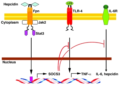
Hepcidin is a peptide hormone that regulates iron homeostasis and acts as an antimicrobial peptide. It is expressed and secreted by a variety of cell types in response to iron loading and inflammation. Hepcidin mediates iron homeostasis by binding to the iron exporter ferroportin, inducing its internalization and degradation via activation of the protein kinase Jak2 and the subsequent phosphorylation of ferroportin. Here we have shown that hepcidin-activated Jak2 also phosphorylates the transcription factor Stat3, resulting in a transcriptional response. Hepcidin treatment of ferroportin-expressing mouse macrophages showed changes in mRNA expression levels of a wide variety of genes. The changes in transcript levels for half of these genes were a direct effect of hepcidin, as shown by cycloheximide insensitivity, and dependent on the presence of Stat3. Hepcidin-mediated transcriptional changes modulated LPS-induced transcription in both cultured macrophages and in vivo mouse models, as demonstrated by suppression of IL-6 and TNF-alpha transcript and secreted protein. Hepcidin-mediated transcription in mice also suppressed toxicity and morbidity due to single doses of LPS, poly(I:C), and turpentine, which is used to model chronic inflammatory disease. Most notably, we demonstrated that hepcidin pretreatment protected mice from a lethal dose of LPS and that hepcidin-knockout mice could be rescued from LPS toxicity by injection of hepcidin. The results of our study suggest a new function for hepcidin in modulating acute inflammatory responses.


Figures

Figure 1
Figure 1. Changes in macrophage mRNA in response to hepcidin.
Mouse bone marrow macrophages were incubated with FAC (10 μM Fe) for 24 hours to induce the expression of Fpn. Cells were then incubated for 4 hours in the absence or presence of hepcidin at a final concentration 1 μg/ml. Cells were solubilized, and RNA extraction was performed. mRNA levels were analyzed by Affymetrix microarray, as described in Methods. The data show the results of 2 independent experiments. A linear scatter plot of gene expression was generated, with upregulated genes labeled in red, downregulated genes in green, and unchanged genes in gray. The boxes denote genes whose transcripts were examined by RT-PCR.
Figure 2
Figure 2. Hepcidin induces a transcriptional response in macrophages.
(A) Mouse bone marrow macrophages were incubated with FAC (10 μM Fe) to induce the expression of Fpn. After 24 hours, cells were incubated in the presence or absence of 1 μg/ml hepcidin for 30 minutes. Cells were placed at 0°C and solubilized and samples immunoprecipitated with rabbit anti-Fpn antibodies. Immunoprecipitated samples were analyzed by Western blot probing for Jak2, Stat3, p-Stat3, or Fpn. (B) Mouse bone marrow macrophages were incubated with FAC (10 μM Fe) for 24 hours, followed by a 4 hour incubation in the absence (–Hep) or presence (+Hep) of hepcidin, Hep20, or protegrin, all at a final concentration of 1 μg/ml. Cells were solubilized, RNA extracted, and Il7, Il17r, and Socs3 mRNAs were analyzed relative to actin mRNA by RT-PCR. (C) Mouse bone marrow macrophages were transfected with either nonspecific (N.S.) or mouse Fpn-, Jak2-, or Stat3-specific siRNA oligonucleotide pools using OligofectAMINE. Cells silenced for mouse Fpn, Jak2, or Stat3 were transfected with siRNA-resistant zebrafish Fpn (Z-Fpn), human Jak2 (H-Jak2), or human Stat3 (H-Stat3), respectively, using Amaxa Nucleofector. Twenty-four hours later, cells were incubated with FAC (10 μM Fe) for 24 hours and then incubated for an additional 4 hours in the absence or presence of hepcidin. Cells were solubilized, RNA was extracted, and Il7 and Il17r mRNA was analyzed relative to actin mRNA by RT-PCR. (D) Mouse bone marrow macrophages were incubated for 24 hours with FAC (10 μM Fe) and then incubated from 0 to 24 hours in the presence of 1 μg/ml hepcidin. Cells were solubilized, RNA was extracted, and Il7, Il17r, and Socs3 mRNA were analyzed relative to actin mRNA by RT-PCR.
Figure 3
Figure 3. Hepcidin modulates inflammatory effects due to LPS.
(A) Mouse bone marrow macrophages were incubated with FAC (10 μM Fe) for 24 hours and then incubated for 2 hours in the absence or presence of hepcidin (1 μg/ml). After 2 hours with or without hepcidin (*), LPS was added for an additional 4 hours, cells were solubilized, and RNA was extracted. Tnfa and Il6 mRNA was analyzed by RT-PCR. (B) Mouse bone marrow macrophages were transfected with either nonspecific or mouse SOCS3-specific siRNA oligonucleotide pools using OligofectAMINE. Forty-eight hours later, cells were incubated in the absence or presence of hepcidin for 2 hours (*) and then incubated with LPS for an additional 4 hours. Cells were solubilized, RNA was extracted, and Il6 and Tnfa mRNA was analyzed relative to actin mRNA by RT-PCR.
Figure 4
Figure 4. Hepcidin suppresses the inflammatory effects of endotoxin in mice.
(A) Age- and sex-matched C57BL/6 mice (5 for each group) were treated with an intraperitoneal injection of PBS or hepcidin (100 μg/mouse). After 2 hours a sublethal dose of LPS (1 mg/kg) was injected intraperitoneally. After 4 hours mice were sacrificed, and mRNA was extracted from liver. mRNA levels were measured for Il6 and Tnfa relative to actin mRNA using RT-PCR. (B) Age- and sex-matched C57BL/6 mice (5 for each group) were treated with an intraperitoneal injection of PBS or hepcidin (100 μg/mouse). After 2 hours a lethal dose of LPS (10 mg/kg) was injected intraperitoneally, morbidity was assessed, and the data were plotted as a Kaplan-Meier plot. (C) C57BL/6 and Hamp–/– mice (5 for each group) were treated with hepcidin (100 μg) or PBS as in A prior to injection of a lethal dose of LPS (10 mg/kg). Mouse morbidity was determined following LPS injection, and the data were plotted using Kaplan-Meier analysis. (D) Wild-type and Hamp–/– mice (5 for each group) were injected with LPS (10 mg/kg), mouse morbidity was determined, and the data were plotted using Kaplan-Meier analysis.
Figure 5
Figure 5. Effect of a high-iron diet on LPS-injected wild-type and Hamp–/– mice.
(A) Six C57BL/6 and (B) 6 Hamp–/– mice maintained on a high-iron diet for 2 days were given an intraperitoneal injection of PBS or LPS (1 mg/kg). After 4 hours mice were sacrificed, and mRNA was extracted from liver and spleen. Liver mRNAs for Tnfa, Il6, Il7, Il17r, and Sos3 were analyzed by RT-PCR. (C) Five C57BL/6 mice were treated with an intraperitoneal injection of PBS or hepcidin (100 μg/mouse). Two hours after hepcidin/PBS injection, mice were given an intraperitoneal injection of PBS, poly(I:C) (100 μg/mouse), or turpentine (100 μl/mouse). After 4 hours mice were sacrificed, sera isolated, and TNF-α and IL-6 (pg/ml serum) levels were measured. (D) Fifteen C57BL/6 mice were subjected to CLP and treated with hepcidin or saline, and IL-6 levels (pg/ml serum) were assayed. *P = 0.039, **P = 0.068, ***P = 0.015. (E) Survival of the mice treated in D was determined. The survival data were plotted as a Kaplan-Meier plot.
Figure 6
Figure 6. Model for hepcidin’s role in modifying inflammation.
LPS binding to TLR-4 results in increased transcripts for inflammatory cytokines such as TNF-α and IL-6. Activation of TLR-4 directly or through IL-6 binding to its receptor induces transcription of hepcidin. Increased serum hepcidin binds to Fpn, resulting in binding and activation of Jak2/Stat3. Activation of Jak2 leads to downregulation of Fpn and decreased iron export into plasma. Activation of Stat3 leads to a transcriptional response resulting in increased concentrations of SOCS3. SOCS3 dampens signal transduction by TLRs and cytokine receptors, resulting in decreased inflammatory cytokines and hepcidin transcription.

Comment in

References

    1. Nemeth E, et al. Hepcidin regulates cellular iron efflux by binding to ferroportin and inducing its internalization. Science. 2004;306(5704):2090–2093. doi: 10.1126/science.1104742. - DOI - PubMed
    1. Park CH, Valore EV, Waring AJ, Ganz T. Hepcidin, a urinary antimicrobial peptide synthesized in the liver. J Biol Chem. 2001;276(11):7806–7810. doi: 10.1074/jbc.M008922200. - DOI - PubMed
    1. Shi J, Camus AC. Hepcidins in amphibians and fishes: Antimicrobial peptides or iron-regulatory hormones? Dev Comp Immunol. 2006;30(9):746–755. - PubMed
    1. Lee PL, Beutler E. Regulation of hepcidin and iron-overload disease. Annu Rev Pathol. 2009;4:489–515. doi: 10.1146/annurev.pathol.4.110807.092205. - DOI - PubMed
    1. De Domenico I, et al. The molecular mechanism of hepcidin-mediated ferroportin down-regulation. Mol Biol Cell. 2007;18(7):2569–2578. doi: 10.1091/mbc.E07-01-0060. - DOI - PMC - PubMed

Publication types

MeSH terms