. 2010 Jul 31;42(7):503-13.
doi: 10.3858/emm.2010.42.7.051.

2-Deoxy-D-glucose regulates dedifferentiation through beta-catenin pathway in rabbit articular chondrocytes

Affiliations

2-Deoxy-D-glucose regulates dedifferentiation through beta-catenin pathway in rabbit articular chondrocytes

Seon Mi Yu et al. Exp Mol Med. .

Abstract

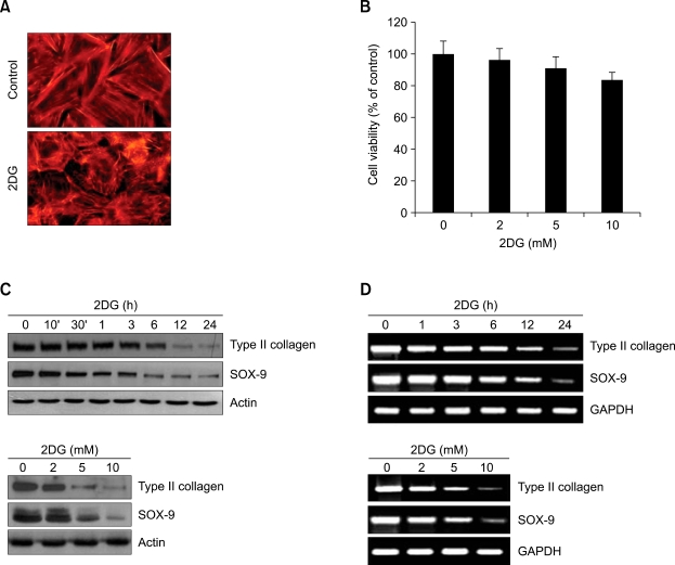
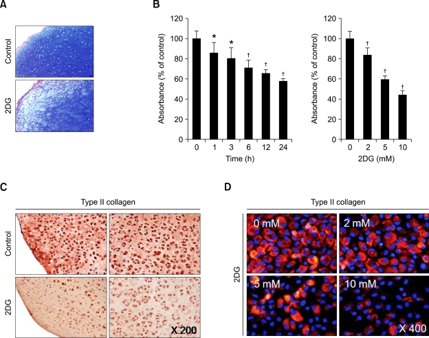
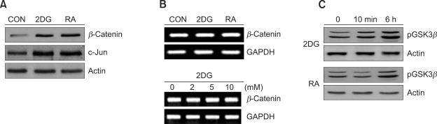
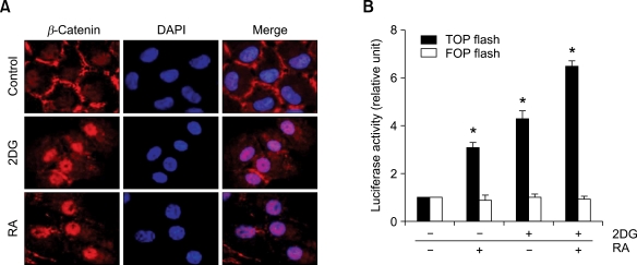
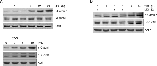
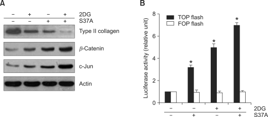
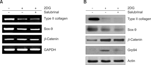
2-deoxy-D-glucose (2DG) is known as a synthetic inhibitor of glucose. 2DG regulates various cellular responses including proliferation, apoptosis and differentiation by regulation of glucose metabolism in cancer cells. However, the effects of 2DG in normal cells, including chondrocytes, are not clear yet. We examined the effects of 2DG on dedifferentiation with a focus on the beta-catenin pathway in rabbit articular chondrocytes. The rabbit articular chondrocytes were treated with 5 mM 2DG for the indicated time periods or with various concentrations of 2DG for 24 h, and the expression of type II collagen, c-jun and beta-catenin was determined by Western blot, RT-PCR, immunofluorescence staining and immunohistochemical staining and reduction of sulfated proteoglycan synthesis detected by Alcain blue staining. Luciferase assay using a TCF (T cell factor)/LEF (lymphoid enhancer factor) reporter construct was used to demonstrate the transcriptional activity of beta-catenin. We found that 2DG treatment caused a decrease of type II collagen expression. 2DG induced dedifferentiation was dependent on activation of beta-catenin, as the 2DG stimulated accumulation of beta-catenin, which is characterized by translocation of beta-catenin into the nucleus determined by immunofluorescence staining and luciferase assay. Inhibition of beta-catenin degradation by inhibition of glycogen synthase kinase 3-beta with lithium chloride (LiCl) or inhibition of proteasome with z-Leu-Leu-Leu-CHO (MG132) accelerated the decrease of type II collagen expression in the chondrocytes. 2DG regulated the post-translational level of beta-catenin whereas the transcriptional level of beta-catenin was not altered. These results collectively showed that 2DG regulates dedifferentiation via beta-catenin pathway in rabbit articular chondrocytes.


Figures

Figure 1
Figure 1
2DG causes dedifferentiation in rabbit articular chondrocytes. (A) Rabbit articular chondrocytes were untreated or treated with 5 mM 2DG for 24 h. Cells were stained for F-actin with rhodamine-conjugated phalloidin. (B) Cells were untreated or treated with the 5 mM 2DG for the indicated time periods. Cell viability was determined by MTT assay. (C) Rabbit articular chondrocytes were untreated or treated with 5 mM 2DG for the indicated time periods (upper panel) or with the specific concentrations of 2DG for 24 h (lower panel). Expressions of type II collagen and SOX-9 were analyzed by Western blot analysis. Expressions of β-actin were used as loading controls. (D) Articular chondrocytes were untreated or treated with 5 mM 2DG for the indicated time periods (upper panel) or with the specific concentrations of 2DG for 24 h (lower panel). Expressions of type II collagen and SOX-9 were detected by RT-PCR. Expressions of glyceraldehyde-3-phosphate dehydrogenase (GAPDH) were used as loading controls. The data represent a typical experiment, whereby similar results were obtained from four independent experiments.
Figure 2
Figure 2
2DG reduces sulfated proteoglycan accumulation and type II collagen expression in cartilage explants and chondrocytes. (A) Articular cartilage was untreated or treated with 5 mM 2DG for 24 h. Accumulation of sulfated proteoglycan was determined by Alcian blue staining of cross-sections of cartilage. (B) Rabbit articular chondrocytes were untreated or treated with 5 mM 2DG for the indicated time periods (left panel) or with the specific concentrations of 2DG for 24 h (right panel). (C) Cartilage explants were untreated or treated with 5 mM 2DG for 24 h or with the specific concentrations of 2DG for 24 h. (D) Primary rabbit articular chondrocytes were untreated or treated with 5 mM 2DG for 24 h or with the specific concentrations of 2DG for 24 h. Cells were stained with DAPI. Type II collagen was determined by immunohistochemical staining (C) and immunofluorescence staining (D). Data are presented as results of a typical experiment (A, C, D) and as mean values with standard deviation (B) (n = 4). *, P < 0.05; , P < 0.01 compared with untreated cells.
Figure 3
Figure 3
2DG stimulates β-catenin expression in rabbit articular chondrocytes. (A) Articular chondrocytes were untreated or treated with 5 mM 2DG or with 1 µM RA for 24 h. Expressions of β-catenin, c-jun and actin were determined by Western blot analysis. (B) Primary chondrocytes were untreated or treated with 5 mM 2DG or with 1 µM RA for 24 h or with the specific concentrations of 2DG for 24 h. Expressions of β-catenin and GAPDH were detected by RT-PCR. GAPDH was used as a loading control. (C) Cells were untreated or treated with 5 mM 2DG or with 1 µM RA for the indicated time periods. Expressions of phosphorylated GSK-3β and actin were analyzed by Western blot analysis. (A, C) Expressions of Actin were used as loading controls of blots. The data represent a typical experiment, whereby similar results were obtained from four independent experiments.
Figure 4
Figure 4
2DG increases nuclear localization of β-catenin in rabbit articular chondrocytes. (A) Articular chondrocytes were untreated or treated with 5 mM 2DG, 1 µM RA, and 5 mM 2DG in the presence of 1 µM RA for 24 h. Expressions of β-catenin and 4,6-diamidini-2-phenylindole (DAPI) were double stained and analyzed by immunofluorescence microscopy. (B) Chondrocytes were transfected with active (TOPFlash) or inactive (FOPFlash) TCF/LEF reporter gene for β-catenin and untreated or treated with 5 mM 2DG or with 1 µM RA or with 5 mM 2DG in the presence of 1 µM RA for 24 h. The TCF/LEF reporter activity was detected using luminometer. Data are presented as results of a typical experiment (A) and as mean values with standard deviation (B) (n = 4). *, P < 0.01 compared with untreated cells.
Figure 5
Figure 5
2DG induces β-catenin expression via pGSK3β in rabbit articular chondrocytes. (A) Articular chondrocytes were untreated or treated with 5 mM 2DG for the indicated time periods (upper panel) or with the specific concentrations of 2DG for 24 h (lower panel). Expressions of β-catenin, phosphorylated GSK-3β and actin were detected by Western blot analysis. (B) Chondrocytes were untreated or treated with 5 mM 2DG for the indicated time periods in the presence of 40 µM proteasome inhibitor Z-Leu-Leu-Leu-al (MG132). Expressions of β-catenin, phosphorylated GSK-3β and actin were detected by Western blot analysis. Data are presented as results of a typical experiment.
Figure 6
Figure 6
Accumulation of β-catenin induces dedifferentiation in rabbit articular chondrocytes. (A) Chondrocytes were untreated or treated with 5 mM 2DG for 24 h in the presence of 10 mM lithium chloride (LiCl) or 40 µM proteasomal inhibitor Z-Leu-Leu-Leu-CHO (MG132). Expressions of type II collagen, β-catenin, c-jun and actin were detected by Western blot analysis. Actin was used as a loading control. (B) Chondrocytes were transfected with active (TOPFlash) or inactive (FOPFlash) TCF/LEF reporter gene and then treated with the vehicle alone as a control (con), 5 mM 2DG or 5 mM 2DG with 10 mM LiCl or 40 µM MG132, and TCF/LEF reporter activity was monitored by luminometer. Data are presented as results of a typical experiment (A) and as mean values with standard deviation (B) (n = 4). *, P < 0.01 compared with untreated cells.
Figure 7
Figure 7
Ectopic expression of S37A β-catenin causes phenotype loss of chondrocytes. (A) Chondrocytes were transfected with empty vector or S37A β-catenin for 6 h, and treated with the vehicle alone as a control (con) or 5 mM 2DG for 24 h. After 24 h incubation, expressions of type II collagen, β-catenin, c-Jun and β-actin were detected by Western blot analysis. Actin was used as loading control. (B) Chondrocytes were transfected with active (TOPFlash) or transfected with inactive (FOPFlash) TCF/LEF reporter gene and S37A β-catenin, and treated with the vehicle alone as a control or with 5 mM 2DG, and TCF/LEF reporter activity was monitored by luminometer. Data are presented as results of a typical experiment (A) and as mean values with standard deviation (B) (n = 4). *, P < 0.01 compared with untreated cells.
Figure 8
Figure 8
Dedifferentiation by 2DG occurs through β-catenin accumulation in rabbit articular chondrocytes. (A) Chondrocytes were untreated or treated with 5 mM 2DG for 24 h in the presence of 20 µM ER-stress inhibitor salubrinal. Expressions of type II collagen, SOX-9, β-catenin and GAPDH were determined by RT-PCR. GAPDH was used as a loading control. (B) Primary chondrocytes were untreated or treated with 5 mM 2DG for 24 h in the presence of 20 µM salubrinal, ER-stress inhibitor. Expressions of type II collagen, SOX-9, β-catenin, Grp94 and β-actin were determined by Western blot analysis. Data are presented as results of a typical experiment.

References

    1. Barth AI, Pollack AL, Altschuler Y, Mostov KE, Nelson WJ. NH2-terminal deletion of beta-catenin results in stable colocalization of mutant beta-catenin with adenomatous polyposis coli protein and altered MDCK cell adhesion. J Cell Biol. 1997;136:693–706. - PMC - PubMed
    1. Blom AB, Brockbank SM, van Lent PL, van Beuningen HM, Geurts J, Takahashi N, van der Kraan PM, van de Loo FA, Schreurs BW, Clements K, Newham P, van den Berg WB. Involvement of the Wnt signaling pathway in experimental and human osteoarthritis: prominent role of Wnt-induced signaling protein 1. Arthritis Rheum. 2009;60:501–512. - PubMed
    1. Cash DE, Bock CB, Schughart K, Linney E, Underhill TM. Retinoic acid receptor alpha function in vertebrate limb skeletogenesis: a modulator of chondrogenesis. J Cell Biol. 1997;136:445–457. - PMC - PubMed
    1. Charni-Ben Tabassi N, Garnero P. Monitoring cartilage turnover. Curr Rheumatol Rep. 2007;9:16–24. - PubMed
    1. Cho SH, Oh CD, Kim SJ, Kim IC, Chun JS. Retinoic acid inhibits chondrogenesis of mesenchymal cells by sustaining expression of N-cadherin and its associated proteins. J Cell Biochem. 2003;89:837–847. - PubMed

Publication types

MeSH terms

LinkOut - more resources