. 2010 Aug 5;29(31):4460-72.
doi: 10.1038/onc.2010.199. Epub 2010 Jun 7.

A mosaic mouse model of astrocytoma identifies alphavbeta8 integrin as a negative regulator of tumor angiogenesis

Affiliations

A mosaic mouse model of astrocytoma identifies alphavbeta8 integrin as a negative regulator of tumor angiogenesis

J H Tchaicha et al. Oncogene. .

Abstract

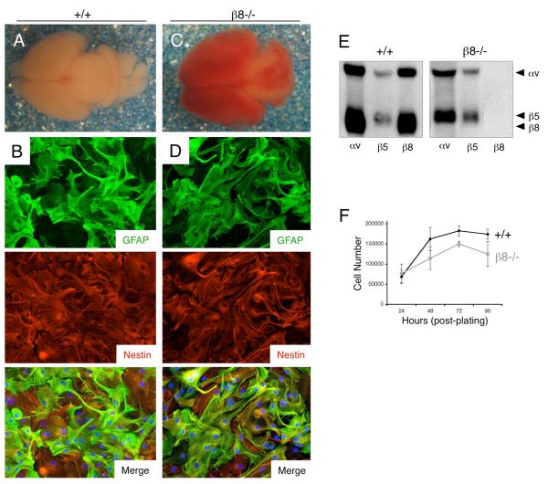
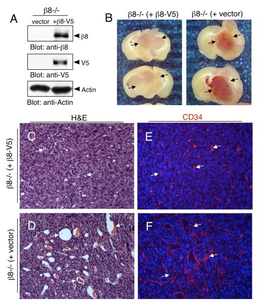
Angiogenesis involves a complex set of cell-cell and cell-extracellular matrix (ECM) interactions that coordinately promote and inhibit blood vessel growth and sprouting. Although many factors that promote angiogenesis have been characterized, the identities and mechanisms of action of endogenous inhibitors of angiogenesis remain unclear. Furthermore, little is known about how cancer cells selectively circumvent the actions of these inhibitors to promote pathological angiogenesis, a requisite event for tumor progression. Using mosaic mouse models of the malignant brain cancer, astrocytoma, we report that tumor cells induce pathological angiogenesis by suppressing expression of the ECM protein receptor alphavbeta8 integrin. Diminished integrin expression in astrocytoma cells leads to reduced activation of latent TGFbetas, resulting in impaired TGFbeta receptor signaling in tumor-associated endothelial cells. These data reveal that astrocytoma cells manipulate their angiogenic balance by selectively suppressing alphavbeta8 integrin expression and function. Finally, these results show that an adhesion and signaling axis normally involved in developmental brain angiogenesis is pathologically exploited in adult brain tumors.


Figures

Figure 1
Figure 1. β8 integrin protein is differentially expressed in human astrocytoma cell lines
(A); Detergent-soluble lysates prepared from five human astrocytoma cell lines were immunoblotted with antibodies recognizing αv and β8 integrin proteins. Note that U87 and LN18 cells express low levels of β8 integrin protein, whereas LN229, SNB19 and U251 cell express robust levels of β8 integrin protein. (B, C); U87 glioma cells, which express low levels of β8 integrin, were stereotactically implanted into the striatum of immunocompromised mice (n=5) and coronal tumor sections were analyzed by H&E staining (B) or anti-CD34 immunohistochemistry (C). Note that U87 cells form large intracranial tumors that are well vascularized and contain hemorrhagic and sinusoidal-like vessels (arrows in C). Boxed areas in B (100X) are shown at higher magnification (400X) in panel C. (D, E); SNB19 glioma cells, which express robust levels of β8 integrin protein, were stereotactically implanted into the striatum of immunocompromised mice (n=3) and coronal tumor sections were analyzed by H&E staining (D) or anti-CD34 immunohistochemistry (E). SNB19 tumors formed in periventricular regions of the brain. These focal lesions contained blood vessels (arrows in E) that are morphologically distinct from blood vessels in U87 tumors (arrows in C). Boxed areas in D (100X) are shown at higher magnification (400X) in panel E.
Figure 2
Figure 2. Mouse astroglial progenitor cells, a presumptive cell type of origin for astrocytomas, express robust levels of endogenous αvβ8 integrin
(A, C); Images of whole brains dissected from wild type (A) and β8−/− (C) newborn littermates. Note the hemorrhage throughout the β8 integrin knockout brain. (B, D); Primary astroglial progenitor cells were cultured from cerebral cortices of wild type (B) or β8−/− (D) neonatal mice and immunofluorescently stained with antibodies recognizing GFAP (upper panels) and nestin (middle panels). Note that most cells co-express GFAP and Nestin protein (lower panels in B, D). (E); Analysis of integrin protein expression in primary astroglial progenitors. Wild type and β8−/− astroglial progenitor cells cultured from neonatal brains were incubated with amine-reactive biotin to label cell surface proteins. Lysates were immunoprecipitated with antibodies directed against αv, β5 or β8 integrins. Note the absence of αvβ8 integrin expression in β8−/− cells. (F); Primary β8−/− astroglial progenitor cells do not display obvious growth differences in vitro, as compared to wild type controls. Cell proliferation under adherent conditions was quantified by counting viable cells every 24 hours over a period of four days (n=3 per genotype per time point). Error bars represent standard deviations.
Figure 3
Figure 3. Integrin-independent cell growth in transformed astroglial progenitor cells
(A, B); Immunofluorescence staining reveals that Nestin protein expression is maintained in wild type (A) and β8−/− (B) astroglial progenitor cells that have been transformed with E6/E7 and G12VH-Ras oncogenes. Note the changes in cell morphologies following transformation, as compared to primary cell morphologies shown in Figures 2B and 2D. (C); Detergent-soluble lysates from wild type and β8−/− primary and transformed cells were immunoblotted with antibodies directed against Ras and E7 proteins. (D); Lysates prepared from primary and transformed astroglial progenitor cells were immunoblotted with anti-Nestin, anti-CD133/Prominin-1, anti-GFAP and anti-Actin antibodies. Note the increase in CD133 expression and decrease in GFAP expression in transformed cells. Lysates used for these immunoblots are the same as those analyzed in Figure 4A, therefore the actin immunoblots are the same. (E); Proliferation indices for adherent wild type and β8−/− transformed astroglial progenitors were determined by counting the total numbers of cells every 24 hours over four consecutive days (n=3 samples per genotype per time point). (F); Adhesion-independent cell growth in soft agar was quantified by counting crystal violet-stained colonies (n=3 samples per genotype). The differences in colony numbers formed from wild type and β8−/− cells were not statistically significant. All error bars represent standard deviations.
Figure 4
Figure 4. Reduced αvβ8 integrin protein expression following oncogene-mediated astroglial cell transformation
(A); Detergent-soluble lysates from wild type or β8−/− primary (prim.) and transformed (trans.) astroglial progenitors were immunoblotted with antibodies directed against αv, β1, and β8 integrins. In comparison to primary cells, note the progressive reduction in αv and β8 integrin protein expression in transformed cells. The lower molecular weight band in the αv integrin immunoblots (transformed samples) is likely non-glycosylated integrin protein. (B); Primary cells (upper panels) and transformed cells (lower panels) were analyzed by fluorescent activated cell sorting using an anti-αv integrin antibody conjugated to phycoerythrein (αv-PE). Percentages of primary and transformed cells expressing cell surface αv integrin protein are indicated in red font. (C); Wild type transformed astroglial progenitor cells were propagated over 12 passages. Detergent-soluble lysates (P2 to P12) were then immunoblotted with anti-αv and anti-β8 antibodies. In comparison to unpassaged primary cells (primary), note the progressive reduction in αvβ8 integrin protein expression as transformed cells are passaged.
Figure 5
Figure 5. Severe vascular pathologies and diminished survival in mice harboring β8−/− astrocytomas
(A, B); Mice harboring wild type (A) or β8−/− (B) astrocytomas were cardiac-perfused with fixative and then coronally sliced at one millimeter intervals. Shown are representative brain slices from tumor-bearing mice. Note that obvious hemorrhage in β8−/− tumors (arrows, top right panel). (C); Kaplan-Meier survival plot for mice harboring wild type (n=20) or β8−/− (n=16) astrocytomas, *p<0.001 at 50% survival. (D); A separate cohort of mice harboring wild type and β8−/− astrocytomas were sacrificed at 14 days (n=9 per genotype) and 21 days (n=8 per genotype) after injection. Tumor volumes were quantified in H&E-stained coronal sections by measuring tumor diameters at the largest cross-sectional area; p=0.529 at 14 days (β8−/− samples in comparison to wild type samples); p=0.643 at 21 days (β8−/− samples in comparison to wild type samples). Error bars represent standard deviations.
Figure 6
Figure 6. Histological analyses of blood vessels in wild type and β8−/− astrocytomas
(A, B); Coronal sections from wild type (A) or β8−/− (B) astrocytomas stained with hematoxylin and eosin (H&E) and analyzed microscopically (200X). Note the abnormally dilated intratumoral blood vessels in β8−/− astrocytomas (arrows). (C, D); Coronal sections from wild type (C) and β8−/− (D) astrocytomas were immunostained with an anti-CD34 antibody and visualized (400X) to label vascular endothelial cells (arrows). (E, F); Coronal sections from wild type (E) and β8−/− (F) astrocytomas were immunofluorescently labeled with anti-CD34 to reveal vascular endothelial cells (green) and anti-desmin to reveal vascular pericytes (red). Note that desmin-expressing pericytes are associated with blood vessels within wild type and β8−/− astrocytomas. The boxed areas are shown in higher magnification in the lower right corners of each panel. Magnification for panels E and F, 200X. (G); Quantitation of CD34 expression in astrocytoma sections. Sections were immunofluorescently labeled with anti-CD34, and four randomly selected fields were analyzed from wild type and β8−/− tumors (n=5 mice per tumor genotype). In comparison to wild type tumors there is significantly more CD34 immunofluorescence in β8−/− astrocytomas, *p<0.001 for β8−/− samples compared to wild type controls. Error bars represent standard deviations.
Figure 7
Figure 7. Exogenous expression of β8 integrin rescues astrocytoma-induced vascular pathologies
(A): β8−/− transformed astrocytes were stably transfected with empty vector or a vector expressing human β8 integrin protein tagged at the C-terminus with a V5 epitope (β8-V5). Cell lysates were then immunoblotted with an antibodies directed against β8 integrin (top panel), V5 (middle panel) or actin (bottom panel). (B); Mice injected with β8−/− transformed astrocytes stably transfected with empty vector (right panel) or β8−/− cells stably expressing β8-V5 protein (left panel) were cardiac-perfused with fixative and brains were coronally sliced at one millimeter intervals. Note the hemorrhage in β8−/− (empty vector) tumors that is absent in the tumors expressing β8-V5 protein (arrows). (C, D); Coronal sections from β8−/− (+ β8V5) astrocytomas (C) or β8−/− (+ vector) astrocytomas (D) were stained with hematoxylin and eosin (H&E). Note that the dilated intratumoral blood vessels in β8−/− tumors are diminished in tumors expressing β8-V5 integrin protein (arrows). (E, F); Coronal sections from astrocytomas were immunofluorescently labeled with anti-CD34 to reveal vascular endothelial cells. Note the obvious differences in blood vessel characteristics in the β8−/− +β8-V5 tumors (E) versus β8−/− tumors (F). Sections in panels C-E were analyzed at 200X magnification.
Figure 8
Figure 8. Analysis of β8 integrin-mediated TGFβ activation and signaling
(A); Diminished TGFβ activation in β8−/− cells using a PAI1-lucferase reporter assay. Conditioned media (+/− exogenous LAP-TGFβ1) from wild type (n=3) or β8−/− (n=3) transformed astroglial progenitors was transferred to MLECs stably transfected with PAI1-Luciferase followed by analysis of luciferase activity. Conditioned media from wild type cells pre-treated with LAP-TGFβ1 induced robust luciferase activity; however, note the reduced luciferase activity using conditioned media from β8−/− cells, *p<0.0001 compared to conditioned media without LAP-TGFβ1; **p<0.004 compared to wild type cells; ***p= 0.04 compared to wild type cells. Error bars represent standard deviations. (B, C); Fluorescent images (400X) of coronal sections from β8−/− astrocytomas stably expressing β8-V5 integrin protein (B) or β8−/− astrocytomas transfected with empty vector (C). Sections were immunofluorescently labeled with anti-CD31 (red) to visualize vascular endothelial cells and anti-pSmad3 (green) to monitor canonical TGFβ signaling. Note that most vessels within β8 integrin-expressing tumors contain nuclear pSmad3 (arrows in B), whereas vessels within β8−/− tumors are largely negative for nuclear pSmad3 (arrows in C) although some nuclear pSmad3 is detected (arrowhead in C). Boxed areas in B and C are shown at higher magnification in lower right corners. (D); Mice injected intracranially with β8−/− (+ vector) cells or β8−/− (+ β8V5) cells were sacrificed, tumors were microdissected, and intratumoral endothelial cells were isolated using magnetic beads coated with anti-CD31 antibodies. Cell lysates were then immunoblotted with antibodies recognizing phosphorylated Smad2/3, total Smad2/3, or actin.

Similar articles

Cited by

References

    1. Abe M, Harpel JG, Metz CN, Nunes I, Loskutoff DJ, Rifkin DB. An assay for transforming growth factor-beta using cells transfected with a plasminogen activator inhibitor-1 promoter-luciferase construct. Anal Biochem. 1994;216:276–84. - PubMed
    1. Adams RH, Alitalo K. Molecular regulation of angiogenesis and lymphangiogenesis. Nat Rev Mol Cell Biol. 2007;8:464–78. - PubMed
    1. Alcantara Llaguno S, Chen J, Kwon CH, Jackson EL, Li Y, Burns DK, et al. Malignant astrocytomas originate from neural stem/progenitor cells in a somatic tumor suppressor mouse model. Cancer Cell. 2009;15:45–56. - PMC - PubMed
    1. Bachoo RM, Maher EA, Ligon KL, Sharpless NE, Chan SS, You MJ, et al. Epidermal growth factor receptor and Ink4a/Arf: convergent mechanisms governing terminal differentiation and transformation along the neural stem cell to astrocyte axis. Cancer Cell. 2002;1:269–77. - PubMed
    1. Bao S, Wu Q, McLendon RE, Hao Y, Shi Q, Hjelmeland AB, et al. Glioma stem cells promote radioresistance by preferential activation of the DNA damage response. Nature. 2006a;444:756–60. - PubMed

Publication types

MeSH terms