. 2010 Aug 12;29(32):4599-610.
doi: 10.1038/onc.2010.212. Epub 2010 Jun 7.

Constitutive and chemokine-dependent internalization and recycling of CXCR7 in breast cancer cells to degrade chemokine ligands

Affiliations

Constitutive and chemokine-dependent internalization and recycling of CXCR7 in breast cancer cells to degrade chemokine ligands

K E Luker et al. Oncogene. .

Abstract

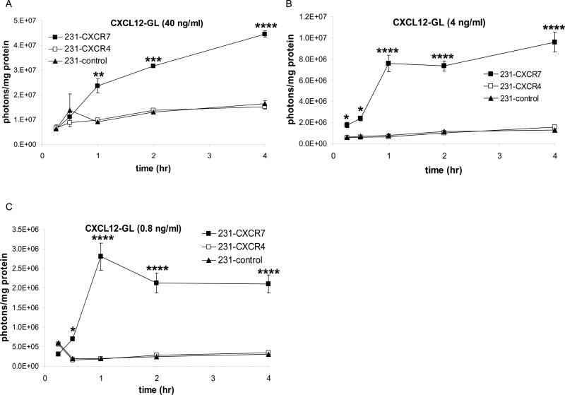
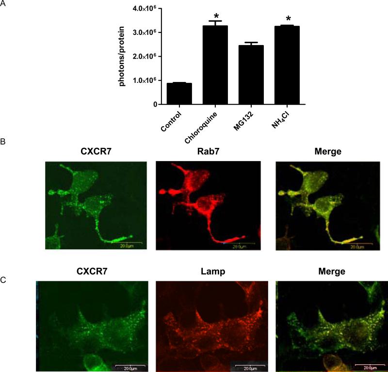
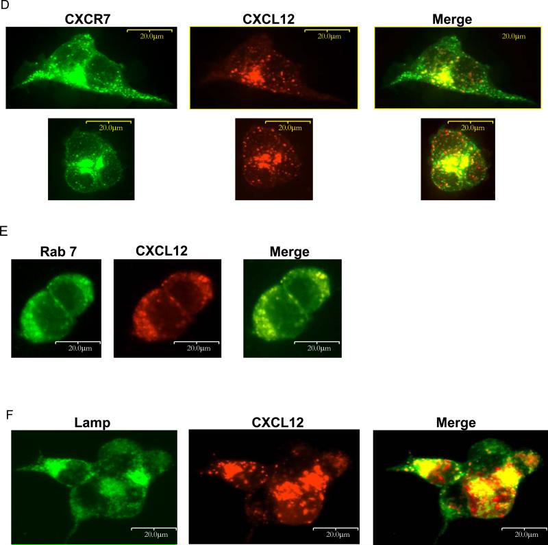
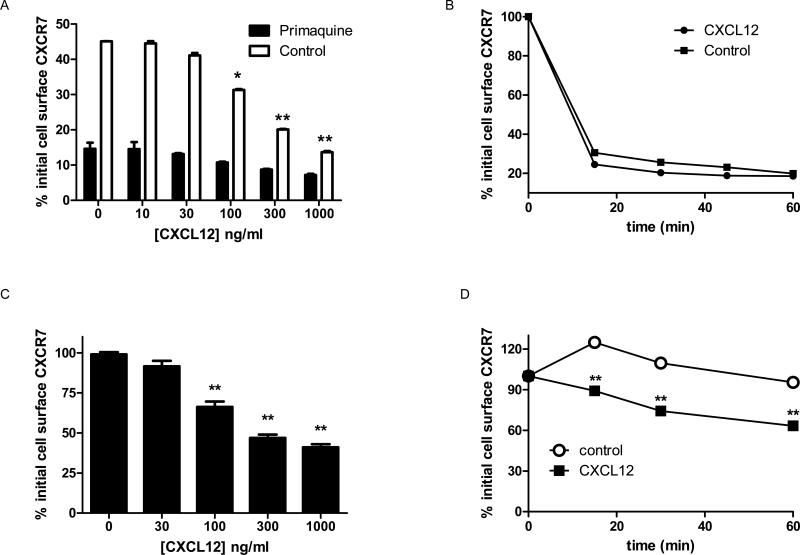
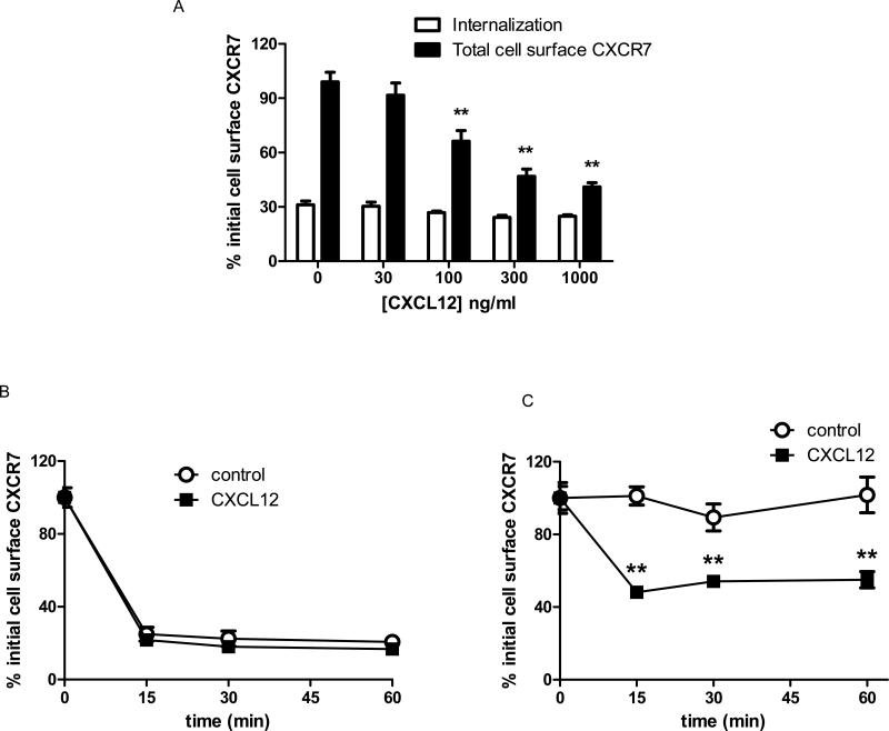
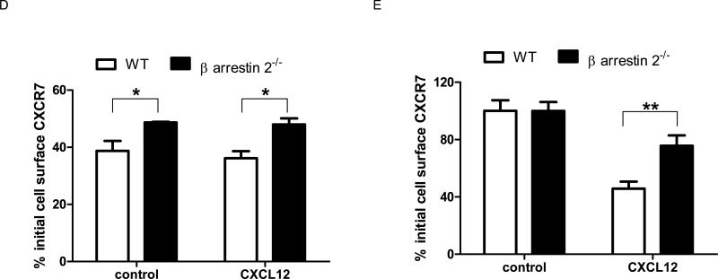
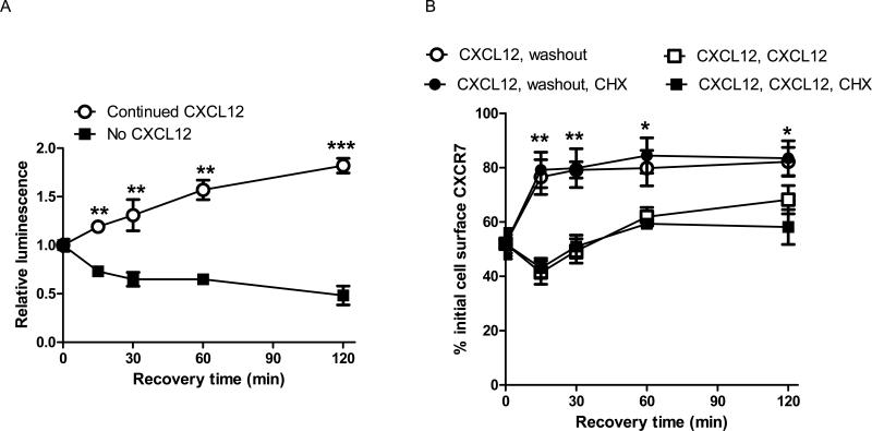
CXCR7 is a receptor for chemokines including CXCL12 (stromal-derived factor-1), a molecule that promotes tumor growth and metastasis in breast cancer and other malignancies. Building on the recent observation that CXCR7 sequesters CXCL12, we investigated mechanisms for CXCR7-dependent uptake of chemokines. Breast cancer cells expressing CXCR7 accumulated chemokines CXCL12 and CXC11 present at concentrations <1 ng/ml, unlike cells expressing CXCR4. CXCR7-dependent accumulation of chemokines was reduced by inhibitors of clathrin-mediated endocytosis. After CXCR7-mediated internalization, CXCL12 trafficked to lysosomes and was degraded, although levels of CXCR7 remained stable. CXCR7 reduced CXCL12 in the extracellular space, limiting the amounts of chemokine available to acutely stimulate signaling through CXCR4. CXCR7 constitutively internalized and recycled to the cell membrane even in the absence of ligand, and addition of chemokines did not significantly enhance receptor internalization. Chemokines at concentrations less than the Kd values for ligand-receptor binding did not alter levels of CXCR7 at the cell surface. Higher concentrations of chemokine ligands reduced the total cell surface expression of CXCR7 without affecting receptor internalization, indicating that receptor recycling was inhibited. CXCR7-dependent uptake of chemokines and receptor trafficking were regulated by beta-arrestin 2. These studies establish mechanisms through which CXCR7 regulates the availability of chemokine ligands in the extracellular space.


Figures

Figure 1
Figure 1. CXCR7-dependent accumulation of CXCL12
MDA-MB-231 cells stably expressing CXCR4 (231-CXCR4), CXCR7 (231-CXCR7), or GFP (231-control) were incubated with ≈ 40 (A), 4 (B), or 0.8 (C) ng/ml CXCL12-GL. Cells were acid washed to remove extracellular chemokine prior to bioluminescence imaging. Data were expressed as mean values of photons/cell protein per well ± SEM (n = 4 each). *, p < 0.05; **, p < 0.01; ***, p < 0.005; ****, p < 0.001.
Figure 2
Figure 2. CXCR7-dependent degradation of chemokines in lysosomes
(A) 231-CXCR7 cells were incubated with ≈ 40 ng/ml CXCL12-GL for 15 minutes, washed, and then incubated without bioluminescent chemokine for 4 hours in medium containing 50 μM chloroquine, 50 mM NH4CL, 25 μM MG132, or vehicle. Cells were acid washed to remove extracellular chemokine before bioluminescence imaging. Photons were normalized to total cellular protein in each sample, and data were expressed as mean values + SEM for per cent initial uptake of CXCL12-GL after the 15 minute pulse (n = 4 each). *, p < 0.05. (B, C) 293T cells were co-transfected with CXCR7-citrine (depicted as green) and either Rab7-CFP (B) or Lamp-CFP (depicted as red) (C), cultured in normal growth medium, and imaged by fluorescence microscopy two days after transfection using a 40X objective. (D – F) 293T cells were transfected with CXCR7-GFP (D) or co-transfected with unfused CXCR7 and Rab7-CFP (E) or Lamp-CFP (F). Two days after transfection, cells were incubated for 30 minutes with ≈ 8 ng/ml CXCL12-cherry. Cells were fixed for fluorescence microscopy and viewed with a 40X objective.
Figure 3
Figure 3. CXCR7 depletes CXCL12 and limits CXCR4 signaling
(A, B) 293T cells were transfected with CXCR7. Localization of CXCR7 and Rab7 (A) 231-CXCR7 or 231-CXCR4 cells were incubated with ≈ 40 ng/ml CXCL12-GL or a comparable amount of unfused GL for 30 minutes and then transferred to the same cell type for an additional 30 minutes. Following two rounds of depletion, bioluminescence from CXCL12-GL and GL was quantified in 5 μl of supernatants. Data were expressed as mean values + SEM for per cent of initial bioluminescence (n = 4 each). (B) 231-CXCR4 cells were incubated with supernatants from B for 5 minutes and then analyzed by Western blot for phosphorylated AKT. Membranes were reprobed for total AKT as a loading control. (C) Supernatants from B were incubated with 293T cells stably expressing a firefly luciferase complementation reporter for activation of CXCR4 (CXCR4-NLuc and β-arrestin 2-CLuc). CXCL12-GL and GL that had not been incubated with 231-CXCR4 or 231-CXCR7 cells were used as controls. Bioluminescence was quantified in live cells, normalized to total protein in each sample, and graphed as mean values + SEM (n = 4 each). *, p < 0.05.
Figure 4
Figure 4. Internalization and cell membrane levels of overexpressed CXCR7
(A) 231-CXCR7 cells were labeled with anti-CXCR7 primary antibody before treatment with increasing concentrations of CXCL12 for 30 minutes to determine receptor internalization in the presence or absence of primaquine to block recycling endosomes. (B) Cells were labeled as in A and then incubated without or with 100 ng/ml CXCL12 in the presence of primaquine. (C) 231-CXCR7 cells were incubated for 30 minutes with increasing concentrations of CXCL12, and then total levels of cell surface CXCR7 were quantified by flow cytometry. (D) Total cell surface CXCR7 was quantified at various times after incubation without or with 100 ng/ml CXCL12. Mean fluorescence intensity for cell surface CXCR7 was measured by flow cytometry, and data were expressed as the percentage of initial cell surface CXCR7. Error bars denote SEM. *, p < 0.05; **, p < 0.01.
Fig 5
Fig 5. Internalization and cell membrane levels of endogenous CXCR7
(A) MCF-7 cells were labeled with anti-CXCR7 antibody and then incubated with various concentrations of CXCL12 for 30 minutes. Receptor internalization and total cell surface levels of CXCR7 at the end of the incubation period were measured by flow cytometry. (B, C) Cells were treated with 100 ng/ml CXCL12 for increasing periods of time before assaying receptor internalization (B) or total cell surface CXCR7 (C) by flow cytometry. Data were expressed as mean values for per cent of initial cell surface CXCR7. *, p < 0.05; **, p < 0.01.
Figure 6
Figure 6. Clathrin-mediated endocytosis and β-arrestin 2 regulate CXCR7-dependent accumulation of chemokines, receptor internalization, and total cell membrane CXCR7
(A) 231-CXCR7 (CXCR7) and 231-control (231) cells were incubated with 0.4M sucrose, 80 μM dynasore, or vehicle control for 30 minutes prior to incubation with ≈ 4 ng/ml CXCL12-GL for various periods of time. Amounts of intracellular chemokine were normalized to total protein per well and graphed as mean ± SEM at each time point (n = 4 per condition). *, p < 0.05 for 231-CXCR7 control samples relative to 231-CXCR7 cells with sucrose or dynasore. (B) β-arrestin 1-/-, β-arrestin 2-/-, and matched wild-type mouse embryonic fibroblasts (MEFs) were transduced with lentiviruses for CXCR7-GFP. Expression of CXCR7-GFP was measured by flow cytometry for GFP. Filled symbols are untransduced cells, while open symbols denote MEFs transduced with CXCR7-GFP. (C) Stably transduced wild-type (WT), β-arrestin 1-/-, or β-arrestin 2-/- MEFs were incubated with ≈ 4 ng/ml CXCL12-GL for 30 minutes in medium containing 100 nM of the CXCR7-specific inhibitor CCX733 or vehicle. Cells were acid washed to remove extracellular chemokine prior to quantifying CXCL12-GL bioluminescence in living cells. Data were graphed as mean values for photons/mg protein + SEM (n = 4 each). (D) Wild-type and β-arrestin 2-/- MEFs were incubated for 30 minutes with 1 mM primaquine in the presence or absence of 100 ng/ml CXCL12 for 30 minutes before quantifying internalization of cell surface CXCR7 by flow cytometry. (E) MEFs were incubated for 30 minutes with or without 100 ng/ml CXCL12, and total mean fluorescence intensity for cell surface CXCR7 at the end of the incubation period was quantified by flow cytometry. Data in D and E were expressed as per cent initial cell surface CXCR7 as in Figure 2. *, p < 0.05; **, p < 0.0.
Figure 7
Figure 7. Removing CXCL12 dissociates CXCR7 from β-arrestin 2 and enhances recovery of cell surface receptors
(A) 231 CXCR7/β-arrestin complementation reporter cells were incubated with 300 ng/ml CXCL12 for 30 minutes (time 0). Parallel samples of cells were washed and then incubated in medium without or with 300 ng/ml CXCL12 for various periods of time. Firefly luciferase activity produced by association of CXCR7 with β-arrestin 2 was quantified by bioluminescence imaging of living cells. Bioluminescence was normalized to total cell protein per well and expressed as changes in luminescence relative to cells at time 0 with no recovery period. Data points are mean values ± SEM (n = 4 per sample). (B) 231-CXCR7 cells were incubated for 30 minutes with 300 ng/ml CXCL12. Cells then were either maintained in CXCL12 or washed to remove extracellular CXCL12 and transferred to fresh medium. Parallel samples of cells were incubated with 100 μg/ml cycloheximide to block protein synthesis during the recovery time. Cell surface CXCR7 was determined using mean values for fluorescence measured by flow cytometry. Error bars denote SEM. *, p < 0.05; **, p < 0.01, ***, p < 0.005.

Similar articles

Cited by

References

    1. Allinen M, R B, Cai L, Brennan C, Lahti-Domerci J, Huang H, et al. Molecular characterization of the tumor microenvironment in breast cancer. Cancer Cell. 2004;6:17–32. - PubMed
    1. Atanackovic D, Cao Y, Kim J, Brandl S, Thorn I, Faltz C, et al. The local cytokine and chemokine milieu within malignant effusions. Tumour Biol. 2008;29:93–104. - PubMed
    1. Balabanian K, Lagane B, Infantino S, Chow K, Harriague J, Moepps B, et al. The chemokine SDF-1/CXCL12 binds to and signals through the orphan receptor RDC1 in T lymphocytes. J Biol Chem. 2005;280:35760–35766. - PubMed
    1. Balkwill F. Cancer and the chemokine network. Nat Rev Cancer. 2004;4:540–550. - PubMed
    1. Boldajipour B, Mahabaleshwar S, Kardash E, Reichman-Fried M, Blaser H, Minina S, et al. Control of chemokine-guided cell migration by ligand sequestration. Cell. 2008;132:463–473. - PubMed

Publication types

MeSH terms