. 2010 Jul 21;29(14):2395-406.
doi: 10.1038/emboj.2010.118. Epub 2010 Jun 8.

PLK2 phosphorylation is critical for CPAP function in procentriole formation during the centrosome cycle

Affiliations

PLK2 phosphorylation is critical for CPAP function in procentriole formation during the centrosome cycle

Jaerak Chang et al. EMBO J. .

Abstract

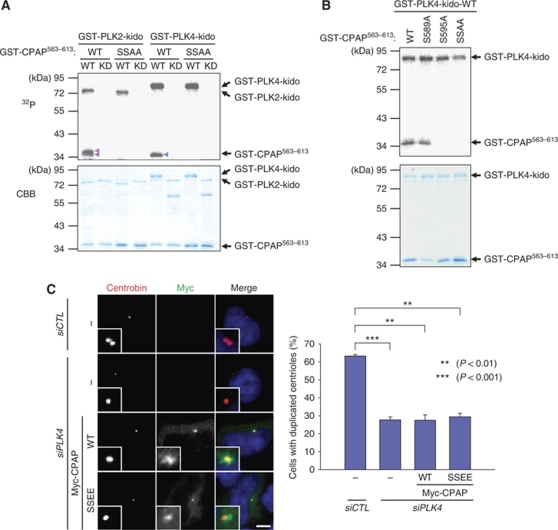
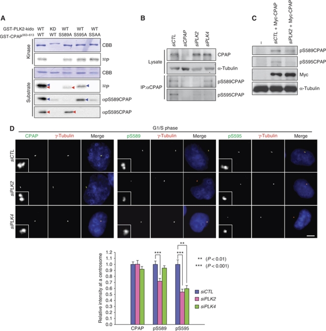
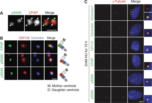
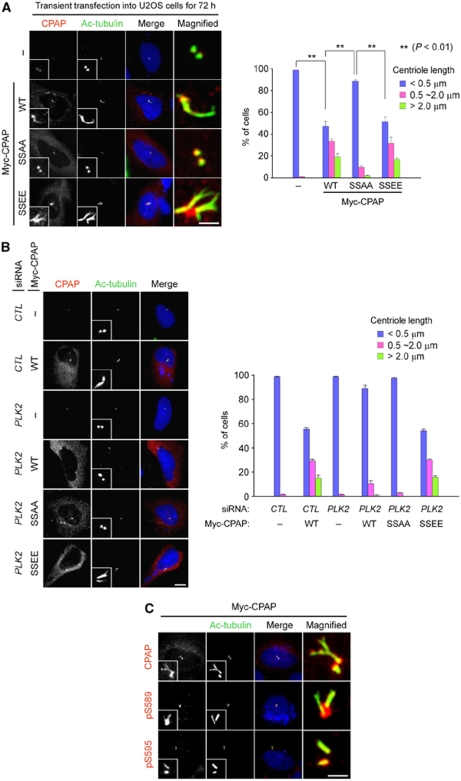
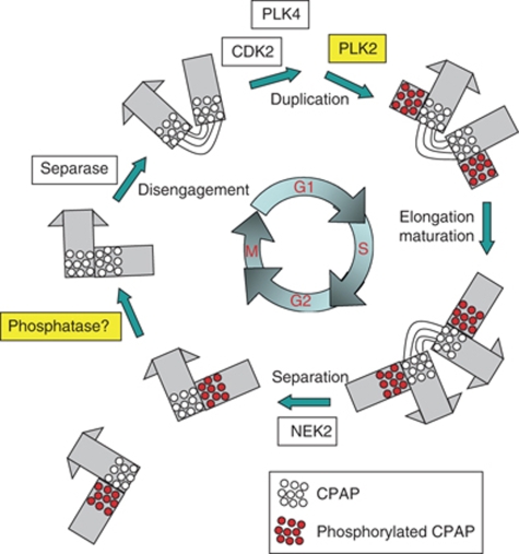
Control of centrosome duplication is tightly linked with the progression of the cell cycle. Recent studies suggest that the fundamental process of centriole duplication is evolutionally conserved. Here, we identified centrosomal P4.1-associated protein (CPAP), a human homologue of SAS-4, as a substrate of PLK2 whose activity oscillates during the cell cycle. PLK2 phosphorylates the S589 and S595 residues of CPAP in vitro and in vivo. This phosphorylation is critical for procentriole formation during the centrosome cycle. PLK4 also phosphorylates S595 of CPAP, but PLK4 phosphorylation is not a critical step in the PLK4 function in procentriole assembly. CPAP is phosphorylated in a cell cycle stage-specific manner, so that its phosphorylation increases at the G1/S transition phase and decreases during the exit of mitosis. Phosphorylated CPAP is preferentially located at the procentriole. Furthermore, overexpression of a phospho-resistant CPAP mutant resulted in the failure to form elongated centrioles. On the basis of these results, we propose that phosphorylated CPAP is involved in procentriole assembly, possibly for centriole elongation. This work demonstrates an example of how procentriole formation is linked to the progression of the cell cycle.


Conflict of interest statement

The authors declare that they have no conflict of interest.

Figures

Figure 1
Figure 1
Phosphorylation of CPAP S589 and S595 is critical for centriole duplication. (A) A summary of the in vitro kinase assays that are presented in Supplementary Figure S1. In vitro kinase assays of PLK2 were performed to determine specific phosphorylation sites within the CPAP protein. As substrates, CPAP was truncated into small pieces. The red lines indicate the truncated CPAP proteins that were strongly phosphorylated by PLK2. The relative phosphorylation intensities are indicated in the right panel. (B) In vitro kinase assays of PLK2 were performed with the mutated GST-CPAP563–613 proteins. The wild type (WT) or kinase dead (KD) forms of GST-PLK21–390-kido (kinase domain) were used for kinases. The phosphorylation activity was examined by autoradiogram. The amounts of the proteins were determined by Coomassie blue staining. (C) In vitro kinase assays of PLK2 were performed with WT or point mutants (S589A, S595A, and S589A/S595A (SSAA)) of GST-CPAP563–613. The red and blue arrowheads indicate the phosphorylation of S589 and S595, respectively. (D) HeLa cells were transfected with pMyc-CPAP or pMyc-CPAPSSAA. Forty-eight hours later, the cells were coimmunostained with antibodies specific to Myc (red) and CP110 (green), and centriole duplication was determined by counting the CP110 dots. Where, 1 or 2 CP110 dot(s), unduplicated centrioles; and 3 or 4 CP110 dots, duplicated centrioles. (E) HeLa cells were transfected with pMyc-CPAP, pMyc-CPAPS589A, pMyc-CPAPS595A, or pMyc-CPAPSSAA. Forty-eight hours later, the cells were coimmunostained with antibodies specific to Myc (green) and centrobin (red), and centriole duplication was determined by counting the centrobin dots. Where, 0 or 1 centrobin dot, unduplicated centrioles; and 2 or 3 centrobin dots, duplicated centrioles. (F) HeLa cells were transfected with siCTL or siCPAP-U and cultured for 48 h. The cell lysates were then subjected to immunoblot analysis with antibodies specific to CPAP and α-tubulin. (G) The siCTL- or siCPAP-U-transfected HeLa cells were coimmunostained with antibodies specific to CPAP (green) and γ-tubulin (red). (H) HeLa cells were transfected with siCTL or siCPAP-U and subsequently with pMyc-CPAP or pMyc-CPAPSSAA. The cells were coimmunostained with antibodies specific to Myc (red) and CP110 (green) to determine centriole duplication. The scale bar represents 10 μm. For statistical analysis, over 50 cells were counted and the experiments were repeated three times. The results are presented as means and standard errors. *P<0.05, analysed with the paired t-test. Insets are magnified views of the centrosomes.
Figure 2
Figure 2
PLK2 phosphorylation of CPAP is essential for centriole duplication. (A) HeLa cells were transfected with either siCTL or siPLK2 and cultured for 72 h. The lysates were subjected to immunoblot analysis with antibodies specific to PLK2 and α-tubulin. (B) The siCTL- or siPLK2-transfected U2OS cells were coimmunostained with antibodies specific to PLK2 (green) and γ-tubulin (red). (C) The siCTL- or siPLK2-transfected HeLa cells were coimmunostained with antibodies specific to CP110 (green) and γ-tubulin (red). The centrioles in the interphase cells were counted. (D) HeLa cells were transfected with siRNAs (siCTL or siPLK2) and subsequently with the CPAP constructs (pMyc-CPAP, pMyc-CPAPS589E, pMyc-CPAPS595E, or pMyc-CPAPSSEE). The cells were coimmunostained with antibodies specific to Myc and centrobin, and centriole duplication was determined. The scale bar represents 10 μm. For statistical analysis, over 50 cells were counted and the experiments were repeated three times. The results are presented as means and standard errors. *P<0.05; **P<0.01; ***P<0.001, analysed with the paired t-test. Insets are magnified views of the centrosomes.
Figure 3
Figure 3
CPAP is also phosphorylated by PLK4. (A) In vitro kinase assays of PLK2 and PLK4 were performed with the mutated GST-CPAP563–613 proteins. The wild type (WT) and kinase dead (KD) forms of GST-PLK21–390-kido (kinase domain) or GST-PLK41–390-kido were used for kinases. The phosphorylation activity was determined by autoradiogram. The amounts of the proteins were determined by Coomassie blue staining. Blue and red arrowheads indicate the S589 and S595 phosphorylated bands, respectively. (B) In vitro kinase assays of PLK4 were performed with the WT or phospho-resistant mutants (S589A, S595A, and S589A/S595A (SSAA)) of GST-CPAP563–613. (C) HeLa cells were transfected with siPLK4 and subsequently with pMyc-CPAP or pMyc-CPAPSSEE. Forty-eight hours later, the cells were coimmunostained with antibodies specific to Myc (green) and centrobin (red), and centriole duplication was determined. The scale bar represents 10 μm. For statistical analysis, over 50 cells were counted and the experiments were repeated three times. The results are presented as means and standard errors. **P<0.01; ***P<0.001, analysed with the paired t-test. Insets are magnified views of the centrosomes.
Figure 4
Figure 4
CPAP phosphorylation in vivo. (A) In vitro kinase assay of PLK2 was performed with GST-CPAP563−613 point mutants (S589A, S595A, and SSAA) as substrates. The kinase activity of PLK2 was determined with an autoradiogram. The same blot was subjected to immunoblot analysis with antibodies specific to phospho-S589 (αpS589CPAP) or phospho-S595 (αpS595CPAP) of CPAP. Blue and red arrowheads indicate the S589 and S595 phosphorylated bands, respectively. (B) HeLa cells were transfected with siCTL, siCPAP, siPLK2 or siPLK4. Total lysates were immunoprecipitated with the CPAP antiserum to concentrate the endogenous CPAP proteins, followed by immunoblotting with the phospho-CPAP antibodies. One hundredth of the total lysates were subjected to immunoblot analysis with CPAP and α-tubulin antibodies, (C) HeLa cells were transfected with siRNAs (siCTL or siPLK2) and subsequently with the Myc-CPAP. The lysates were subjected to immunoblot analysis with phospho-CPAP (pS589CPAP and pS595CPAP), Myc and α-tubulin antibodies. (D) HeLa cells were transfected with siCTL, siPLK2, or siPLK4. The cells were then synchronized at G1/S phase by a double thymidine block. The cells were immunostained with the CPAP or phospho-CPAP (pS589CPAP and pS595CPAP) antibodies (green) and with the γ-tubulin antibody (red). Immunostaining intensities of the centrosomal CPAP and phospho-CPAP proteins were determined densitometrically relative to the intensity of siCTL-transfected cells. The scale bar represents 10 μm. For statistical analysis, immunofluorescent intensities of at least 20 cells were determined, and the results are presented as means and standard errors. **P<0.01; ***P<0.001, analysed with the t-test. Insets are magnified views of the centrosomes.
Figure 5
Figure 5
Cell cycle stage-specific phosphorylation of CPAP. (A) HeLa cells were treated with nocodazole for 14 h to arrest the cell cycle at M phase and transferred to a fresh medium to synchronously resume the cell cycle. At the indicated time points, the cells were fixed and immunostained with the CPAP or phospho-CPAP (pS589CPAP and pS595CPAP) (green) antibodies, along with the γ-tubulin antibody (red). Immunostaining intensities of the centrosomal CPAP and phospho-CPAP proteins were determined densitometrically relative to the intensity of the 4 h-released cells. Cell cycle synchronization was confirmed by immunoblot analysis of cyclin A and phospho-histone H3 (pHH3). (B) Actively proliferating HeLa cells were immunostained with the CPAP or phospho-CPAP pS589CPAP and pS595CPAP) (green) antibodies and with the γ-tubulin antibody (red). Mitotic phases of the cells were determined by DAPI staining of the chromosomes. The immunostaining intensities of the centrosomal CPAP and phospho-CPAP proteins were determined densitometrically relative to the intensity of the prometaphase cells. Scale bar represents 10 μm. For statistical analysis, immunofluorescent intensities of at least 10 cells were determined and the results are presented as means and standard errors. Insets are magnified views of the centrosomes.
Figure 6
Figure 6
Phosphorylated CPAP localizes to the procentrioles. (A) Centrioles in U2OS cells were coimmunostained with CPAP (red) and pS589CPAP (green) antibodies. Arrows and arrowheads indicate the parental centrioles and procentriole, respectively. The scale bar represents 1 μm. (B) HeLa cells were accumulated at G2 phase by a thymidine block and release for 7 h for detection of the cells with separated centrosomes. The centrioles were triple stained with antibodies specific to CEP135 (red) and centrobin (blue), along with CPAP or phospho-CPAP (pS589CPAP and pS595CPAP, green). The scale bar represents 1 μm. (C) U2OS cells were treated with hydroxyurea (HU) for 72 h and coimmunostained with hCenexin1, centrobin, CP110, CPAP, or phospho-CPAP (pS589CPAP and pS595CPAP) (green) antibodies, along with the γ-tubulin antibody (red). The scale bar represents 10 μm. Insets are magnified views of the centrosomes.
Figure 7
Figure 7
Phosphorylation of CPAP S589 and S595 is required for centriole elongation. (A) U2OS cells were transfected with pMyc-CPAP, pMyc-CPAPSSAA, or pMyc-CPAPSSEE. Seventy-two hours later, the cells were placed on ice to depolymerize unstable microtubules, fixed, and coimmunostained with antibodies specific to CPAP (red) and acetylated tubulin (green). The scale bar represents 2 μm. For statistical analysis, over 100 cells were counted and the experiments were repeated three times. The results are presented as means and standard errors. **P<0.01, analysed with the paired t-test. (B) U2OS cells were transfected with siRNAs (siCTL or siPLK2) and subsequently with the CPAP constructs (pMyc-CPAP, pMyc-CPAPSSAA, or pMyc-CPAPSSEE). The cells were coimmunostained with antibodies specific to CPAP (red) and acetylated tubulin (green). The scale bar represents 10 μm. For statistical analysis, over 50 cells were counted and the experiments were repeated three times. (C) U2OS cells were transfected with pMyc-CPAP. Seventy-two hours later, the cells were coimmunostained with antibodies specific to CPAP or phospho-CPAP (pS589CPAP and pS595CPAP) (red) antibodies, along with the acetylated-tubulin antibody (green). The scale bar represents 2 μm. Insets are magnified views of the centrosomes.
Figure 8
Figure 8
Model. A number of protein kinases control the centriole cycle in a close association with the cell cycle. PLK2 phosphorylation is important for the function of CPAP in procentriole formation at G1/S transition phase. The phosphorylated CPAP (red dots) is located at the proximal end of the procentriole. CPAP is dephosphorylated by an unknown phosphatase once the cell enters anaphase (blank dots).

Similar articles

Cited by

References

    1. Azimzadeh J, Bornens M (2007) Structure and duplication of the centrosome. J Cell Sci 120 (Pt 13): 2139–2142 - PubMed
    1. Bettencourt-Dias M, Glover DM (2007) Centrosome biogenesis and function: centrosomics brings new understanding. Nat Rev Mol Cell Biol 8: 451–463 - PubMed
    1. Bettencourt-Dias M, Rodrigues-Martins A, Carpenter L, Riparbelli M, Lehmann L, Gatt MK, Carmo N, Balloux F, Callaini G, Glover DM (2005) SAK/PLK4 is required for centriole duplication and flagella development. Curr Biol 15: 2199–2207 - PubMed
    1. Bond J, Roberts E, Springell K, Lizarraga SB, Scott S, Higgins J, Hampshire DJ, Morrison EE, Leal GF, Silva EO, Costa SM, Baralle D, Raponi M, Karbani G, Rashid Y, Jafri H, Bennett C, Corry P, Walsh CA, Woods CG (2005) A centrosomal mechanism involving CDK5RAP2 and CENPJ controls brain size. Nat Genet 37: 353–355 - PubMed
    1. Chen Z, Indjeian VB, McManus M, Wang L, Dynlacht BD (2002) CP110, a cell cycle-dependent CDK substrate, regulates centrosome duplication in human cells. Dev Cell 3: 339–350 - PubMed

Publication types

MeSH terms

Substances