. 2010 Jul 21;29(14):2358-67.
doi: 10.1038/emboj.2010.122. Epub 2010 Jun 8.

Dis3-like 1: a novel exoribonuclease associated with the human exosome

Affiliations

Dis3-like 1: a novel exoribonuclease associated with the human exosome

Raymond H J Staals et al. EMBO J. .

Abstract

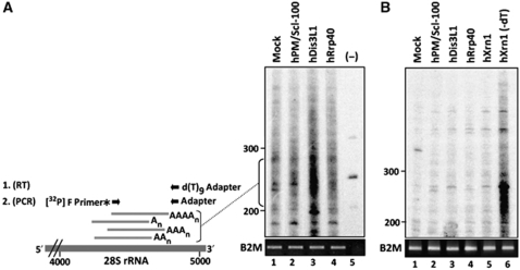
The exosome is an exoribonuclease complex involved in the degradation and maturation of a wide variety of RNAs. The nine-subunit core of the eukaryotic exosome is catalytically inactive and may have an architectural function and mediate substrate binding. In Saccharomyces cerevisiae, the associated Dis3 and Rrp6 provide the exoribonucleolytic activity. The human exosome-associated Rrp6 counterpart contributes to its activity, whereas the human Dis3 protein is not detectably associated with the exosome. Here, a proteomic analysis of immunoaffinity-purified human exosome complexes identified a novel exosome-associated exoribonuclease, human Dis3-like exonuclease 1 (hDis3L1), which was confirmed to associate with the exosome core by co-immunoprecipitation. In contrast to the nuclear localization of Dis3, hDis3L1 exclusively localized to the cytoplasm. The hDis3L1 isolated from transfected cells degraded RNA in an exoribonucleolytic manner, and its RNB domain seemed to mediate this activity. The siRNA-mediated knockdown of hDis3L1 in HeLa cells resulted in elevated levels of poly(A)-tailed 28S rRNA degradation intermediates, indicating the involvement of hDis3L1 in cytoplasmic RNA decay. Taken together, these data indicate that hDis3L1 is a novel exosome-associated exoribonuclease in the cytoplasm of human cells.


Conflict of interest statement

The authors declare that they have no conflict of interest.

Figures

Figure 1
Figure 1
Schematic structure of Dis3 and Dis3-like proteins in yeast and human beings. Schematic diagram of S. cerevisiae Dis3 (yDis3), human Dis3 (hDis3), human Dis3-like 1 (hDis3L1) and human Dis3-like 2 (hDis3L2) with the identified domains indicated: the PIN domain, two cold shock domains (CSD1, CSD2), an RNB domain and an S1 RNA-binding domain. Crackled boxes and question marks indicate domains with a low level of conservation (see Supplementary Figure S1 for details). The level of sequence similarity between the human Dis3 proteins and yDis3 is indicated on the right.
Figure 2
Figure 2
Association of hDis3L1 with the exosome core complex. Total cell extracts were prepared from HEp-2 cells transiently transfected with expression constructs encoding EGFP, EGFP-hDis3L1 or hDis3L1-EGFP, after which immunoprecipitation was performed using polyclonal anti-GFP antibodies. Both the complete cell extracts (Lysate) and the anti-GFP (co-)precipitated proteins (IP) were separated by SDS–PAGE and analysed by western blotting, using monoclonal anti-GFP antibodies and monoclonal anti-hRrp4 antibodies to visualize the co-precipitation of the exosome core complex with hDis3L1. Monoclonal anti-γ-tubulin antibodies were used as loading control.
Figure 3
Figure 3
Localization of hDis3L1 and hDis3 in HEp-2 cells. HEp-2 cells were transiently transfected with expression constructs encoding EGFP-hDis3L1 (A), hDis3L1-EGFP (B), VSV-hDis3L1 (C) or EGFP-hDis3 (D). Forty-eight hours after transfection, the cells were fixed and EGFP-fusion proteins were visualized directly by fluorescence microscopy. The VSV-tagged hDis3L1 protein was visualized by incubating the cells with monoclonal anti-VSV-tag antibodies, followed by Alexa Fluor 555-conjugated goat anti-mouse antibodies and fluorescence microscopy. (EJ) Fixed HEp-2 cells were incubated with polyclonal antibodies to hDis3L1 (FG) or to hDis3 (IJ) and bound antibodies were visualized by Alexa Fluor 488-conjugated secondary antibodies and confocal immunofluorescence microscopy. Nuclei were visualized by Hoechst staining (E, H). (G) and (J) show the merged images of Hoechst and antibody staining. Bar: 10 μm.
Figure 4
Figure 4
In vitro RNA degradation by hDis3L1. (A) HEp-2 cells were transfected with expression constructs encoding EGFP-hDis3L1 or EGFP, and after 48 h, cell lysates were subjected to immunoprecipitation with anti-GFP antibodies. Precipitated proteins/complexes were incubated with a radiolabelled RNA substrate (Input) and the reaction products were subsequently analysed by denaturing polyacrylamide gel electrophoresis followed by autoradiography. The incubations were performed in the presence of increasing concentrations of Mg2+. (B) A similar assay was performed with endogenous exosome complexes after precipitation with anti-hRrp40 antibodies or antibodies from normal rabbit serum (NRS) and lysates from cells, which were transfected with siRNAs downregulating either EGFP (used as a control) or hDis3L1. (C) Expression efficiency of EGFP-tagged hDis3L1 proteins monitored by western blotting using anti-GFP antibodies. WT: wild-type hDis3L1. D62N, D166N, D486N: hDis3L1 amino-acid substitution mutants. Anti-γ-tubulin antibodies were used as a loading control. (D) Activity assay as described above (A) with immunoprecipitated hDis3L1 mutants D62N, D166N, D486N in the presence of 0.05 mM Mg2+. Precipitates from cell lysates containing EGFP and wild-type (WT) hDis3L1 were used as negative and positive control, respectively.
Figure 5
Figure 5
Cytoplasmic accumulation of adenylated degradation intermediates after hDis3L1 knockdown. (A) Cytoplasmic RNA from cells in which the indicated proteins were silenced by RNAi was subjected to oligo(dT) RT–PCR labelling, followed by fractionation by denaturing polyacrylamide gel electrophoresis and autoradiography. A schematic representation of the labelling procedure is shown on the left. First, cDNA was generated using a d(T)9-adapter primer. Products from these reactions were amplified by PCR using a [32P]-labelled forward (F) primer corresponding to a sequence element of the 28S rRNA and a reverse primer corresponding to the adapter sequence. Material from a control reaction, in which the reverse transcriptase was omitted (−), is shown in lane 5. The positions of dsDNA markers are indicated on the left of the gel. For normalization, RT–PCR products obtained with PCR primers specific for the B2M gene are shown in the lower panel. (B) A similar oligo(dT) RT–PCR-labelling experiment as in panel A, but now the isolated RNA was incubated with oligo(dT) and RNase H before the RT reactions. RNA isolated from cells in which hXrn1 was knocked down, which similar to silencing of hDis3L1 leads to accumulation of poly(A) containing degradation intermediates, was used as a control to show that treatment with RNase H in the absence of oligo(dT) (lane 6) did not lead to the disappearance of the intermediates.

Comment in

  • Twins take the job.
    Gas ME, Séraphin B. Gas ME, et al. EMBO J. 2010 Jul 21;29(14):2260-1. doi: 10.1038/emboj.2010.148. EMBO J. 2010. PMID: 20648048 Free PMC article. No abstract available.

Similar articles

Cited by

References

    1. Allmang C, Kufel J, Chanfreau G, Mitchell P, Petfalski E, Tollervey D (1999a) Functions of the exosome in rRNA, snoRNA and snRNA synthesis. EMBO J 18: 5399–5410 - PMC - PubMed
    1. Allmang C, Petfalski E, Podtelejnikov A, Mann M, Tollervey D, Mitchell P (1999b) The yeast exosome and human PM-Scl are related complexes of 3′ → 5′ exonucleases. Genes Dev 13: 2148–2158 - PMC - PubMed
    1. Amblar M, Arraiano CM (2005) A single mutation in Escherichia coli ribonuclease II inactivates the enzyme without affecting RNA binding. FEBS J 272: 363–374 - PubMed
    1. Bonneau F, Basquin J, Ebert J, Lorentzen E, Conti E (2009) The yeast exosome functions as a macromolecular cage to channel RNA substrates for degradation. Cell 139: 547–559 - PubMed
    1. Brouwer R, Allmang C, Raijmakers R, van AY, Vree Egberts W, Petfalski E, van Venrooij WJ, Tollervey D, Pruijn GJ (2001a) Three novel components of the human exosome. J Biol Chem 276: 6177–6184 - PubMed

Publication types

MeSH terms