. 2010 Jul 7;29(13):2182-93.
doi: 10.1038/emboj.2010.111. Epub 2010 Jun 8.

Meiosis requires a translational positive loop where CPEB1 ensues its replacement by CPEB4

Affiliations

Meiosis requires a translational positive loop where CPEB1 ensues its replacement by CPEB4

Ana Igea et al. EMBO J. .

Abstract

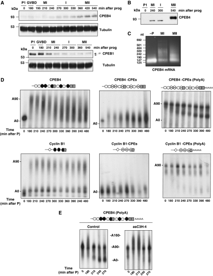
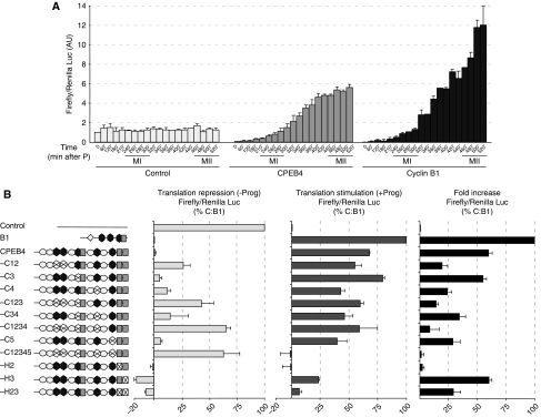
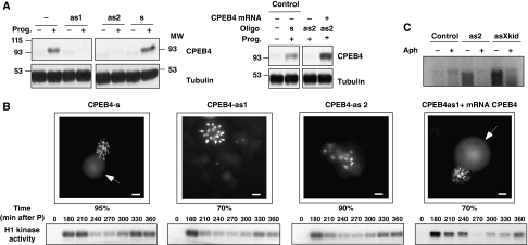
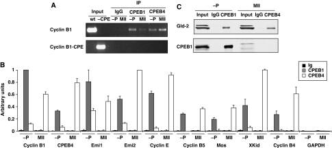
Meiotic progression is driven by the sequential translational activation of maternal messenger RNAs stored in the cytoplasm. This activation is mainly induced by the cytoplasmic elongation of their poly(A) tails, which is mediated by the cytoplasmic polyadenylation element (CPE) present in their 3' untranslated regions. Although polyadenylation in prophase I and metaphase I is mediated by the CPE-binding protein 1 (CPEB1), this protein is degraded during the first meiotic division. Thus, raising the question of how the cytoplasmic polyadenylation required for the second meiotic division is achieved. In this work, we show that CPEB1 generates a positive loop by activating the translation of CPEB4 mRNA, which, in turn, replaces CPEB1 and drives the transition from metaphase I to metaphase II. We further show that CPEB1 and CPEB4 are differentially regulated by phase-specific kinases, generating the need of two sequential CPEB activities to sustain cytoplasmic polyadenylation during all the meiotic phases. Altogether, this work defines a new element in the translational circuit that support an autonomous transition between the two meiotic divisions in the absence of DNA replication.


Conflict of interest statement

The authors declare that they have no conflict of interest.

Figures

Figure 1
Figure 1
CPEB4 mRNA polyadenylation results in CPEB4 accumulation during the second meiotic division. (A) Xenopus oocytes stimulated with progesterone (prog) were collected at the indicated times and analysed by western blotting using anti-CPEB4, anti-CPEB1 or anti-tubulin antibodies. The meiotic phases of the oocyte are indicated (PI, prophase I; GVBD, germinal vesicle breakdown; MI, metaphase I; I, interkinesis; MII, metaphase II). GVBD was determined by the appearance of the white spot at the animal pole of the oocyte. (B) Xenopus oocytes, untreated or stimulated with progesterone (prog), were collected at the indicated times and analysed by western blotting using anti-CPEB4. The meiotic phases of the oocyte are indicated (PI, prophase I; MI, metaphase I; I, interkinesis; MII, metaphase II). (C) Total RNA extracted from oocytes untreated (−P) or incubated with progesterone and collected at metaphase I (MI) and metaphase II (MII) were analysed by RNA-ligation-coupled RT–PCR. (D) Oocytes were injected with the indicated radiolabelled 3′ UTRs. Total RNA was extracted from oocytes collected at the indicated times after progesterone stimulation and analysed by gel electrophoresis followed by autoradiography. Schematic representation of the 3′ UTRs is shown: CPEs as dark grey hexagons, Hexanucleotide as grey boxes, PBEs as rhombus, putative AREs elements as light grey ovals. CPE point mutations are indicated as a cross. (E) Oocytes were injected with C3H-4 anti-sense oligonucleotide (asC3H-4) or C3H-4 sense oligonucleotide (control). After 16 h, oocytes were injected with the indicated radiolabelled 3′ UTRs. Total RNA was extracted from oocytes collected at the indicated times after progesterone stimulation and analysed by gel electrophoresis followed by autoradiography.
Figure 2
Figure 2
CPEB4 is translationally activated by CPEB1 during meiotic maturation. (A, B) The indicated in vitro transcribed Firefly luciferase chimaerical mRNAs were co-injected into oocytes together with Renilla luciferase as a normalization control. (A) Firefly luciferase ORF fused to a control 3′ UTR of 470 nucleotides (control); cyclin B1 3′ UTR wild type (cyclin B1 3′ UTR) and CPEB4 3′ UTR wild type (CPEB4). Oocytes were stimulated with progesterone, collected at the indicated times and the luciferase activities were measured. Data are mean±s.d. (n=4). (B) The indicated Firefly luciferase-3′ UTR variants were injected in oocytes. Oocytes were then incubated in the absence (repression) or presence (activation) of progesterone and the luciferase activities determined after 6 h. The percentage of translational repression in the absence of progesterone (left panel) was normalized to control (100% translation) and to the fully repressed B1 (0% translation). The percentage of translation stimulation (middle panel) was normalized to control (0% stimulation) and B1 (100% simulation). The total fold increase, as the total stimulation by progesterone for each mRNA normalized to control (0% stimulation) and B1 (100% stimulation) is shown in the further right panel. Data are mean±s.d. (n=5). A schematic representation of the 3′ UTR, as in Figure 1, is shown.
Figure 3
Figure 3
CPEB4 synthesis is required for the MI to MII transition. (A, B) Xenopus oocytes were injected with CPEB4 sense (s) or anti-sense (as1, as2) oligonucleotides as indicated and incubated for 16 h. Then, the oocytes were microinjected with CPEB4-enconding mRNA and incubated in the presence or absence of progesterone (prog) as indicated. All the oocytes were collected 4 h after the control, non-injected oocytes, displayed 100% GVBD and analysed as follows. (A) The oocytes were analysed for CPEB4 levels by western blot using anti-CPEB4 and anti-tubulin antibodies (two oocyte equivalents were loaded per lane). (B) Oocytes were fixed, stained with Hoechst and examined under epifluorescence microscope. Representative images and the percentage of appearance for each phenotype are shown. The arrow indicates the first polar body. Scale bar=10 μm. Oocytes collected at the indicated times after progesterone stimulation were analysed for H1 kinase activity as described in Materials and methods. (C) Oocytes injected with CPEB4 anti-sense oligonucleotide (as2), CPEB4 sense oligonucleotide (control) and Xkid anti-sense oligonucleotide (asXkid) were incubated for 16 h and then injected with 0.4 μCi [α-32P]dCTP. Then, the oocytes were stimulated with progesterone and incubated in the presence or absence of Aphydicolin (Aph) as indicated. Oocytes were collected 5 h after control oocytes displayed 100% GVBD, DNA was extracted and analysed by agarose gel electrophoresis followed by autoradiography.
Figure 4
Figure 4
CPEB1 and CPEB4 are sequentially associated with CPE-containing mRNAs. (A) Xenopus oocytes were microinjected with in vitro transcribed RNAs derived from WT cyclin B1 3′ UTR (cyclin B1) or the corresponding variant with the CPEs inactivated by point mutations (cyclin B1-CPE). Then, the oocytes were incubated for 8 h in the presence (MII) or absence (−P) of progesterone and subjected to immunoprecipitation with anti-CPEB1, anti-CPEB4 and control IgG antibodies followed by RT–PCR for the microinjected RNAs. The PCR products derived from the microinjected (input) and co-immunoprecipitated (IP) RNAs were visualized by stained agarose gel electrophoresis. (B, C) Cytoplasmic extracts from oocytes untreated (−P) or incubated with progesterone for 8 h (MII) were subjected to immunoprecipitation with anti-CPEB1, anti-CPEB4 and control IgG antibodies. The co-immunoprecipitates were analysed by qRT–PCR for the presence of the indicated mRNAs (B) or by western blotting for the presence of GLD2 and CPEB1 proteins (C). Data are mean±s.d. (n=3).
Figure 5
Figure 5
A stable CPEB1 mutant cannot replace CPEB4 in the second meiotic division. Xenopus oocytes were injected with CPEB4 sense (control) or anti-sense (as2) oligonucleotides. After 16 h, oocytes were microinjected with mRNAs encoding either CPEB4 or CPEB1-CA and incubated with progesterone. (A) Oocytes were collected at the indicated times and analysed for CPEB1 levels by western blot using anti-CPEB1 and anti-tubulin antibodies (1.5 oocyte equivalents were loaded per lane) (B) Oocytes were collected 4 h after control oocytes display 100% GVBD and treated as Figure 3B. (C, D) Total RNA from oocytes collected at the indicated times was extracted and polyadenylation status of cyclin B1and cyclin E mRNAs was measured by RNA-ligation-coupled RT–PCR.
Figure 6
Figure 6
A stable and constitutively active CPEB1 mutant can compensate for CPEB4 depletion in the second meiotic division. Xenopus oocytes were injected with CPEB4 sense (control) or anti-sense (as2) oligonucleotides. After 16 h, oocytes were microinjected with mRNA encoding a stable and constitutively active CPEB1 mutant (CPEB1-CAD) and incubated with progesterone. Oocytes were collected 4 h after control oocytes display 100% GVBD and analysed for CPEB1 levels by western blot using anti-CPEB1 and anti-tubulin antibodies (one oocyte equivalents were loaded per lane) (A) or fixed, stained with Hoechst and examined under epifluorescence microscope as in Figure 3B (B). (C) Total RNA from oocytes collected at the indicated times was extracted and polyadenylation status of cyclin E and Emi2 mRNAs was measured by RNA-ligation-coupled RT–PCR.
Figure 7
Figure 7
Schematic diagram showing the sequential activities of CPEB1 and CPEB4 mediating the three waves of polyadenylation driving meiotic progression.

Similar articles

Cited by

References

    1. Aoki K, Matsumoto K, Tsujimoto M (2003) Xenopus cold-inducible RNA-binding protein 2 interacts with ElrA, the Xenopus homolog of HuR, and inhibits deadenylation of specific mRNAs. J Biol Chem 278: 48491–48497 - PubMed
    1. Ballantyne S, Daniel DL Jr, Wickens M (1997) A dependent pathway of cytoplasmic polyadenylation reactions linked to cell cycle control by c-mos and CDK1 activation. Mol Biol Cell 8: 1633–1648 - PMC - PubMed
    1. Belloc E, Mendez R (2008) A deadenylation negative feedback mechanism governs meiotic metaphase arrest. Nature 452: 1017–1021 - PubMed
    1. Belloc E, Pique M, Mendez R (2008) Sequential waves of polyadenylation and deadenylation define a translation circuit that drives meiotic progression. Biochem Soc Trans 36(Part 4): 665–670 - PubMed
    1. Charlesworth A, Ridge JA, King LA, MacNicol MC, MacNicol AM (2002) A novel regulatory element determines the timing of Mos mRNA translation during Xenopus oocyte maturation. EMBO J 21: 2798–2806 - PMC - PubMed

Publication types