. 2010 Aug;18(8):1458-61.
doi: 10.1038/mt.2010.106. Epub 2010 Jun 8.

Eight years of clinical improvement in MPTP-lesioned primates after gene therapy with AAV2-hAADC

Affiliations

Eight years of clinical improvement in MPTP-lesioned primates after gene therapy with AAV2-hAADC

Piotr Hadaczek et al. Mol Ther. 2010 Aug.

Abstract

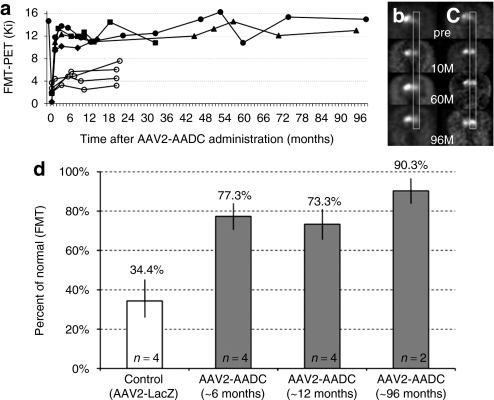
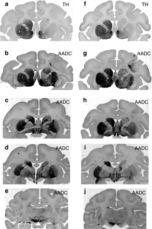
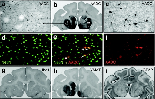
This study completes the longest known in vivo monitoring of adeno-associated virus (AAV)-mediated gene expression in nonhuman primate (NHP) brain. Although six of the eight parkinsonian NHP originally on study have undergone postmortem analysis, as described previously, we monitored the remaining two animals for a total of 8 years. In this study, NHP received AAV2-human L-amino acid decarboxylase (hAADC) infusions into the MPTP (1-methyl-4-phenyl-1,2,3,6-tetrahydropyridine)-lesioned putamen. Restoration of AADC activity restored normal response to levodopa and gene expression could be quantitated repeatedly over many years by 6-[(18)F]fluoro-meta-tyrosine (FMT)-positron emission tomography (PET) and confirm that AADC transgene expression remained unchanged at the 8-year point. Behavioral assessments confirmed continued, normalized response to levodopa (improvement by 35% over historical controls). Postmortem analysis showed that, although only 5.6 + or - 1% and 6.6 + or - 1% of neurons within the transduced volumes of the striatum were transduced, this still secured robust clinical improvement. Importantly, there were no signs of neuroinflammation or reactive gliosis at the 8-year point, indicative of the safety of this treatment. The present data suggest that the improvement in the L-3,4-dihydroxyphenylalanine (L-Dopa) therapeutic window brought about by AADC gene therapy is pronounced and persistent for many years.


Figures

Figure 1
Figure 1
PET imaging of hAADC activity in hemi-lesioned monkey striatum. Data before 72 months are taken from Bankiewicz et al. (a) AADC activity (measured by Ki) over time. Open circles represent four control monkeys that received AAV2-LacZ after MPTP lesioning. Solid symbols indicated AAV2-hAADC-treated NHP; two NHP, Octopus (triangle) and Max (circles), remained in life out to 96 months. (b,c) PET images from AAV2-hAADC-treated animals, (b) Octopus and (c) Max. Images indicate coronal [18F]FMT PET images at baseline (pre) and at 10, 60, and 96 months after unilateral AAV2-hAADC administration to the right side of the brain. Both monkeys showed a stable and long-term increase in AADC activity. (d) Increase in FMT-PET signal after AAV2-AADC treatment over time. This restoration of signal was expressed as a percentage of the control/baseline FMT-PET signal (FMT uptake value in normal monkeys before MPTP lesioning). As there was no significant change in FMT uptake within the control or treated monkeys over time, we grouped animals into three time points (AAV2-AADC-treated monkeys; gray columns). The white column represents the mean FMT-PET signal from control monkeys, MPTP-lesioned monkeys and animals infused with AAV2-LacZ. FMT Ki values and earlier time points for the AAV2-AADC-treated monkeys were calculated from Bankiewicz et al. Standard deviations are shown for each group and the difference between the control and treated animals (at each time point) is statistically significant, P < 0.005. AADC, -amino acid decarboxylase; AAV, adeno-associated virus; FMT, 6-[18F]fluoro-meta-tyrosine; MPTP, 1-methyl-4-phenyl-1,2,3,6-tetrahydropyridine; NHP, nonhuman primate; PET, positron emission tomography.
Figure 2
Figure 2
Extent of AADC transgene distribution within NHP brains. Two monkey's, Max (a–e) and Octopus (f–j) were treated long-term with AAV2-AADC. Consecutive sections (in rows) show precommissural (b,g), commissural (c,h), postcommissural (d,i), and midbrain (e,j) brain regions. The signal in the left hemisphere shows endogenous AADC expression. The right hemispheres show the extent of restoration of AADC expression in the MPTP-lesioned brains. Note the dramatic reduction of anti-TH-staining in the right striatum (a, f) compared to the intact left side, confirming profound loss of dopaminergic fibers and terminals on the lesioned side. AADC, -amino acid decarboxylase; AAV, adeno-associated virus; MPTP, 1-methyl-4-phenyl-1,2,3,6- tetrahydropyridine; TH, tyrosine hydroxylase.
Figure 3
Figure 3
Immunohistochemistry of the brain in hemi-lesioned monkeys 96 months after AAV2-AADC treatment. (a–c) AADC staining in one NHP (Max). The signal shows endogenous AADC in the unlesioned, left striatum, and almost complete restoration of the enzyme activity within the right striatum demonstrating the (b) long-term expression of AADC transgene. Endogenous AADC expression is characterized by (a) dense network of positive fibers whereas the restored lesioned side shows (c) transduced neuronal cell bodies along with the network of their processes. (d–f) Double immunofluorescent labeling of the AAV2-AADC-transduced hemisphere (Max) shows colocalization of markers AADC and NeuN (neuronal marker). The transduced neurons displayed a typical medium spinal morphology. (h) Staining against vesicular monoamine transporter (VMAT2), a marker for presynaptic neurons, shows a dramatic loss of dopaminergic terminals on the MPTP-lesioned, right hemisphere. The left side shows normal expression (compare to similar pattern of TH-staining—Figure 2a). (g) Staining for a microglia marker, Iba1, shows no inflammatory reaction on the AAV2-AADC treated side. Similarly, the lack of astrocytic activation was confirmed with (i) marker GFAP. AADC, -amino acid decarboxylase; AAV, adeno-associated virus; MPTP, 1-methyl-4-phenyl-1,2,3,6-tetrahydropyridine; NHP, nonhuman primate; TH, tyrosine hydroxylase.

References

    1. Edelstein ML, Abedi MR., and, Wixon J. Gene therapy clinical trials worldwide to 2007–an update. J Gene Med. 2007;9:833–842. - PubMed
    1. Fiandaca M, Forsayeth J., and, Bankiewicz K. Current status of gene therapy trials for Parkinson's disease. Exp Neurol. 2008;209:51–57. - PubMed
    1. Forsayeth JR, Eberling JL, Sanftner LM, Zhen Z, Pivirotto P, Bringas J, et al. A dose-ranging study of AAV-hAADC therapy in Parkinsonian monkeys. Mol Ther. 2006;14:571–577. - PMC - PubMed
    1. Bankiewicz KS, Forsayeth J, Eberling JL, Sanchez-Pernaute R, Pivirotto P, Bringas J, et al. Long-term clinical improvement in MPTP-lesioned primates after gene therapy with AAV-hAADC. Mol Ther. 2006;14:564–570. - PubMed
    1. Eberling JL, Jagust WJ, Christine CW, Starr P, Larson P, Bankiewicz KS, et al. Results from a phase I safety trial of hAADC gene therapy for Parkinson disease. Neurology. 2008;70:1980–1983. - PubMed

Publication types

LinkOut - more resources