. 2010 May 28;5(5):e10891.
doi: 10.1371/journal.pone.0010891.

Evidence for a role of endocannabinoids, astrocytes and p38 phosphorylation in the resolution of postoperative pain

Affiliations

Evidence for a role of endocannabinoids, astrocytes and p38 phosphorylation in the resolution of postoperative pain

Matthew S Alkaitis et al. PLoS One. .

Abstract

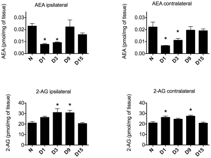
Background: An alarming portion of patients develop persistent or chronic pain following surgical procedures, but the mechanisms underlying the transition from acute to chronic pain states are not fully understood. In general, endocannabinoids (ECBs) inhibit nociceptive processing by stimulating cannabinoid receptors type 1 (CB(1)) and type 2 (CB(2)). We have previously shown that intrathecal administration of a CB(2) receptor agonist reverses both surgical incision-induced behavioral hypersensitivity and associated over-expression of spinal glial markers. We therefore hypothesized that endocannabinoid signaling promotes the resolution of acute postoperative pain by modulating pro-inflammatory signaling in spinal cord glial cells.

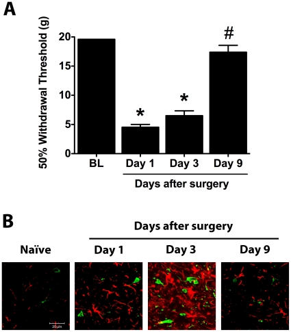
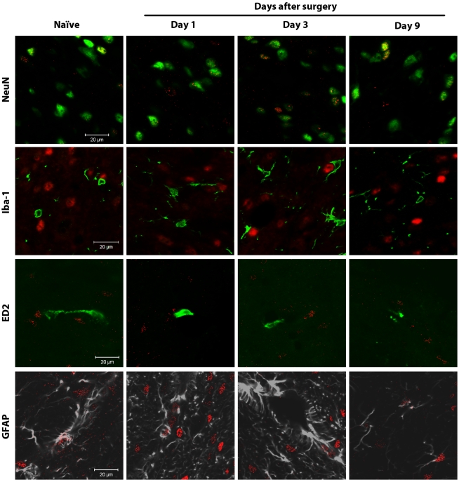
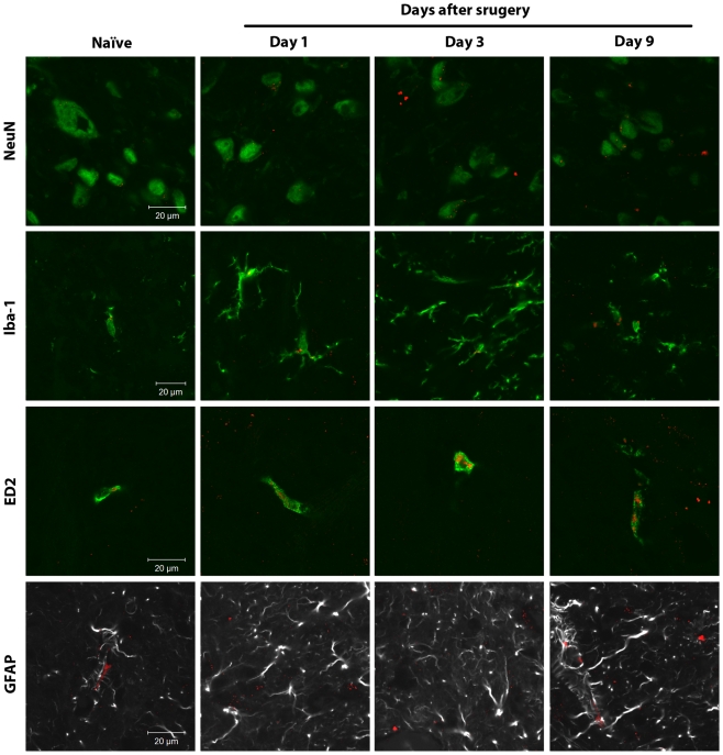
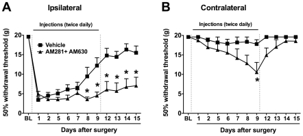
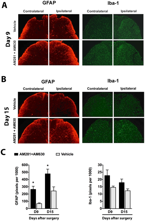
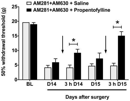
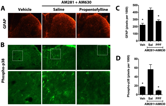
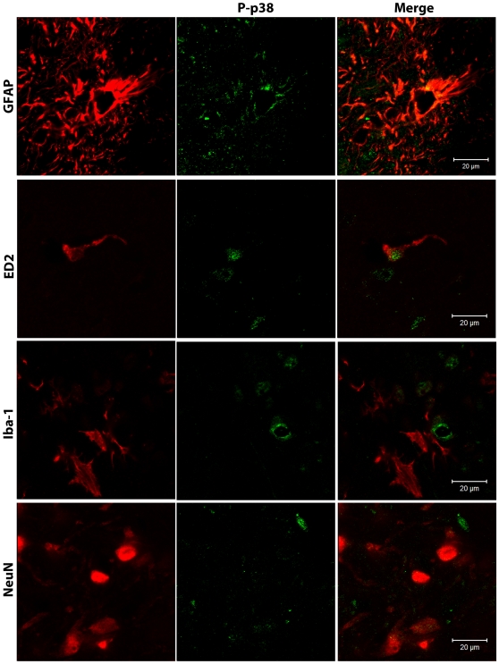
Methodology/principal findings: To test this hypothesis, rats receiving paw incision surgery were used as a model of acute postoperative pain that spontaneously resolves. We first characterized the concentration of ECBs and localization of CB(1) and CB(2) receptors in the spinal cord following paw incision. We then administered concomitant CB(1) and CB(2) receptor antagonists/inverse agonists (AM281 and AM630, 1 mg x kg(-1) each, i.p.) during the acute phase of paw incision-induced mechanical allodynia and evaluated the expression of glial cell markers and phosphorylated p38 (a MAPK associated with inflammation) in the lumbar dorsal horn. Dual blockade of CB(1) and CB(2) receptor signaling prevented the resolution of postoperative allodynia and resulted in persistent over-expression of spinal Glial Fibrillary Acidic Protein (GFAP, an astrocytic marker) and phospho-p38 in astrocytes. We provide evidence for the functional significance of these astrocytic changes by demonstrating that intrathecal administration of propentofylline (50 microg, i.t.) attenuated both persistent behavioral hypersensitivity and over-expression of GFAP and phospho-p38 in antagonist-treated animals.

Conclusions/significance: Our results demonstrate that endocannabinoid signaling via CB(1) and CB(2) receptors is necessary for the resolution of paw incision-induced behavioral hypersensitivity and for the limitation of pro-inflammatory signaling in astrocytes following surgical insult. Our findings suggest that therapeutic strategies designed to enhance endocannabinoid signaling may prevent patients from developing persistent or chronic pain states following surgery.


Conflict of interest statement

Competing Interests: The American Pain Society 2008 Future Leaders in Pain Research Small Grant (AR-S) does not alter the authors' adherence to all the PLoS ONE policies on sharing data and materials.

Figures

Figure 1
Figure 1. Paw incision-induced mechanical allodynia and increased glial marker expression spontaneously resolve.
(A) 50% paw withdrawal thresholds were evaluated in naïve animals and ipsilateral to paw incision in animals at days 1, 3 and 9 after surgery (n = 8 per group). *p<0.05 vs. baseline values by one-way ANOVA repeated measures followed by Dunnett's post test. #p<0.05 vs. postoperative day 1 values by one-way ANOVA repeated measures followed by Dunnett's post test. (B) L5 spinal cord sections were stained for Iba-1 (green) and GFAP (red). Representative confocal images show detail of the superficial laminae of the L5 dorsal horn in naïve animals and ipsilateral to paw incision in rats at days 1, 3 and 9 after surgery. BL: Baseline.
Figure 2
Figure 2. Spinal endocannabinoid levels are dynamically regulated following paw incision.
Spinal cord concentrations of AEA and 2-AG were determined in the lumbar spinal cord of naïve rats (N, n = 3) and in the ipsilateral and contralateral lumbar spinal cord of rats at days 1 (D1), 3 (D3), 9 (D9) and 15 (D15) after paw incision surgery (n = 6 for each group). *p<0.05 vs. naïve group by one-way ANOVA followed by Dunnett's post test. 2-AG: 2-Arachidonoylglycerol, AEA: Anandamide.
Figure 3
Figure 3. Spinal CB1 receptors are mainly expressed in neurons.
Confocal analysis was used to determine spinal CB1 receptor cellular localization in the superficial laminae of the L5 dorsal horn in naïve rats or ipsilateral to surgery in rats at days 1, 3 and 9 after paw incision. Representative images are shown. CB1 receptor staining appears in red. NeuN (marker for neurons), Iba-1 (marker for microglia) and ED2/CD163 (ED2, marker for perivascular microglia) appear in green. GFAP (marker for astrocytes) appears in grey. In the images of CB1 receptors and Iba-1, CB1 receptor staining originally appeared green, and Iba-1 appeared in red. These colors were digitally switched in order to consistently represent CB1 receptor staining in red in all images. In the images of CB1 receptors and GFAP, GFAP staining originally appeared in green. The color of GFAP staining was digitally changed to grey in order to allow better visualization of occasional staining of CB1 receptors on GFAP-positive cells. The colocalization of CB1 receptors and NeuN staining appears in yellow.
Figure 4
Figure 4. Spinal CB2 receptors are mainly expressed in microglial cells.
Confocal analysis was used to determine CB2 receptor cellular localization in the superficial laminae of the L5 dorsal horn in naïve rats or ipsilateral to surgery in rats at days 1, 3 and 9 after paw incision. Representative images are shown. CB2 receptor staining appears in red. NeuN (marker for neurons), Iba-1 (marker for microglia) and ED2/CD163 (ED2, marker for perivascular microglia) appear in green. GFAP (marker for astrocytes) appears in grey. In the images of CB2 receptors and GFAP, GFAP staining originally appeared in green. The color of GFAP staining was digitally changed to grey in order to allow a better visualization of this specific marker and any potential staining of CB2 receptors. The colocalization of CB2 receptors with the other cellular markers is visualized in yellow.
Figure 5
Figure 5. Dual blockade of CB1 and CB2 receptors prevents the normal resolution of paw incision-induced hypersensitivity.
Withdrawal thresholds ipsilateral (A) and contralateral (B) to paw incision were determined before (BL: Baseline) and following surgery. Mixed antagonists of CB1 receptors (AM281; 1 mg.kg−1) and CB2 receptors (AM630; 1 mg.kg−1) or vehicle were administered twice daily (i.p.) through day 9 (doted vertical lines). *p<0.05 vs. vehicle group by two-way ANOVA repeated measures followed by Bonferroni post test. n = 8 per group.
Figure 6
Figure 6. Dual blockade of CB1 and CB2 receptors results in persistent over-expression of GFAP.
Representative images show GFAP (astrocytic marker) and Iba-1 (microglial marker) staining in the L5 dorsal horn ipsilateral and contralateral to paw incision in vehicle- and AM281 + AM630-treated rats at postoperative days 9 (A) and 15 (B). Quantification of these markers in laminae I-II is shown in the bottom panel (C). Staining was quantified as the number of pixels above a set threshold per total pixels in the selected area. Vehicle group: Iba-1-day 9, n = 4; Iba-1-day 15, n = 8; GFAP-day 9, n = 3; GFAP-day 15, n = 8. AM281+AM630 group: Iba-1-day 9, n = 4; Iba-1-day 15, n = 6; GFAP-day 9, n = 3; GFAP-day 15, n = 6. *p<0.05 vs. vehicle group by two-way ANOVA followed by Bonferroni post test.
Figure 7
Figure 7. Propentofylline reverses behavioral hypersensitivity in rats treated with dual CB1/CB2 receptor blockade.
50% withdrawal thresholds were determined in AM281 + AM630-treated animals in response to subsequent propentofylline or saline treatment (indicated by arrows). Animals treated from days 1-9 with mixed CB antagonists were treated on days 14 and 15 with the glial modulator propentofylline (50 µg in 10 µl, i.t., n = 7) or saline (10 µl, i.t., n = 7). Behavior was tested before and 3 hours (3h) after injections as shown. *p<0.05 vs. saline group by two-way ANOVA repeated measures followed by Bonferroni post tests. BL: Baseline.
Figure 8
Figure 8. Propentofylline reverses over-expression of GFAP and phospho-p38 in rats treated with dual CB1/CB2 receptor blockade.
Representative images show spinal GFAP (A) and phospho-p38 staining (B) in the ipsilateral L5 dorsal horn of animals treated from days 1-9 with AM281 + AM630 and subsequently treated on days 14 and 15 with the glial modulator propentofylline (PPF, 50 µg in 10 µl, i.t., n = 4) or saline (Sal, 10 µl, i.t., n = 3). Staining of GFAP (C) and phospho-p-38 (D) was quantified as the number of pixels above a set threshold per total pixels in the selected area. Controls (Veh, n = 3) were treated with the antagonist vehicle for nine days but did not receive either propentofylline or saline treatment. *p<0.05 vs. AM281 + AM630/saline group by one-way ANOVA followed by Dunnett's post test.
Figure 9
Figure 9. Phospho-p38 is expressed in astrocytes and perivascular microglia.
Confocal analysis was used to determine phospho-p38 (P-p38) cellular localization in the superficial laminae of the ipsilateral L5 dorsal horn in AM281 + AM630-treated rats on day 15 after paw incision. Representative images are shown. Phospho-p38 appears in green. GFAP (marker for astrocytes), ED2/CD163 (ED2, marker for perivascular microglia), Iba-1 (marker for microglia) and NeuN (marker for neurons) appear in red. The color of ED2/CD163, Iba-1 and NeuN staining was digitally changed from green to red, and phospho-p38 from red to green for consistency in presenting the data and in order to allow a better visualization of the co-localization of these markers. The colors of the GFAP/phospho-p38 co-stain (top panel) were not altered.

Similar articles

Cited by

References

    1. Kehlet H, Jensen TS, Woolf CJ. Persistent postsurgical pain: risk factors and prevention. Lancet. 2006;367:1618–1625. - PubMed
    1. Guindon J, Hohmann AG. The endocannabinoid system and pain. CNS Neurol Disord Drug Targets. 2009;8:403–421. - PMC - PubMed
    1. Romero-Sandoval A, Eisenach JC. Spinal cannabinoid receptor type 2 activation reduces hypersensitivity and spinal cord glial activation after paw incision. Anesthesiology. 2007;106:787–794. - PubMed
    1. Romero-Sandoval A, Nutile-McMenemy N, DeLeo JA. Spinal microglial and perivascular cell cannabinoid receptor type 2 activation reduces behavioral hypersensitivity without tolerance after peripheral nerve injury. Anesthesiology. 2008;108:722–734. - PMC - PubMed
    1. Scott DA, Wright CE, Angus JA. Evidence that CB-1 and CB-2 cannabinoid receptors mediate antinociception in neuropathic pain in the rat. Pain. 2004;109:124–131. - PubMed

Publication types

MeSH terms