The acid test of fluoride: how pH modulates toxicity
- PMID: 20531944
- PMCID: PMC2878349
- DOI: 10.1371/journal.pone.0010895
The acid test of fluoride: how pH modulates toxicity
Abstract
Background: It is not known why the ameloblasts responsible for dental enamel formation are uniquely sensitive to fluoride (F(-)). Herein, we present a novel theory with supporting data to show that the low pH environment of maturating stage ameloblasts enhances their sensitivity to a given dose of F(-). Enamel formation is initiated in a neutral pH environment (secretory stage); however, the pH can fall to below 6.0 as most of the mineral precipitates (maturation stage). Low pH can facilitate entry of F(-) into cells. Here, we asked if F(-) was more toxic at low pH, as measured by increased cell stress and decreased cell function.
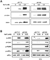
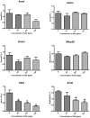
Methodology/principal findings: Treatment of ameloblast-derived LS8 cells with F(-) at low pH reduced the threshold dose of F(-) required to phosphorylate stress-related proteins, PERK, eIF2alpha, JNK and c-jun. To assess protein secretion, LS8 cells were stably transduced with a secreted reporter, Gaussia luciferase, and secretion was quantified as a function of F(-) dose and pH. Luciferase secretion significantly decreased within 2 hr of F(-) treatment at low pH versus neutral pH, indicating increased functional toxicity. Rats given 100 ppm F(-) in their drinking water exhibited increased stress-mediated phosphorylation of eIF2alpha in maturation stage ameloblasts (pH<6.0) as compared to secretory stage ameloblasts (pH approximately 7.2). Intriguingly, F(-)-treated rats demonstrated a striking decrease in transcripts expressed during the maturation stage of enamel development (Klk4 and Amtn). In contrast, the expression of secretory stage genes, AmelX, Ambn, Enam and Mmp20, was unaffected.
Conclusions: The low pH environment of maturation stage ameloblasts facilitates the uptake of F(-), causing increased cell stress that compromises ameloblast function, resulting in dental fluorosis.
Conflict of interest statement
Figures


Similar articles
-
Sirtuin1 and autophagy protect cells from fluoride-induced cell stress.Biochim Biophys Acta. 2014 Feb;1842(2):245-55. doi: 10.1016/j.bbadis.2013.11.023. Epub 2013 Dec 1. Biochim Biophys Acta. 2014. PMID: 24296261 Free PMC article.
-
High amounts of fluoride induce apoptosis/cell death in matured ameloblast-like LS8 cells by downregulating Bcl-2.Arch Oral Biol. 2013 Sep;58(9):1165-73. doi: 10.1016/j.archoralbio.2013.03.016. Epub 2013 Apr 15. Arch Oral Biol. 2013. PMID: 23598055
-
Magnesium, pH regulation and modulation by mouse ameloblasts exposed to fluoride.Bone. 2017 Jan;94:56-64. doi: 10.1016/j.bone.2016.10.014. Epub 2016 Oct 13. Bone. 2017. PMID: 27744011
-
Function and repair of dental enamel - Potential role of epithelial transport processes of ameloblasts.Pancreatology. 2015 Jul;15(4 Suppl):S55-60. doi: 10.1016/j.pan.2015.01.012. Epub 2015 Feb 14. Pancreatology. 2015. PMID: 25747281 Review.
-
The impact of fluoride on ameloblasts and the mechanisms of enamel fluorosis.J Dent Res. 2009 Oct;88(10):877-93. doi: 10.1177/0022034509343280. J Dent Res. 2009. PMID: 19783795 Free PMC article. Review.
Cited by
-
How pH is regulated during amelogenesis in dental fluorosis.Exp Ther Med. 2018 Nov;16(5):3759-3765. doi: 10.3892/etm.2018.6728. Epub 2018 Sep 11. Exp Ther Med. 2018. PMID: 30402142 Free PMC article. Review.
-
Fluoride does not inhibit enamel protease activity.J Dent Res. 2011 Apr;90(4):489-94. doi: 10.1177/0022034510390043. Epub 2010 Nov 30. J Dent Res. 2011. PMID: 21118795 Free PMC article.
-
Assessment of dental fluorosis in Mmp20 +/- mice.J Dent Res. 2011 Jun;90(6):788-92. doi: 10.1177/0022034511398868. Epub 2011 Mar 8. J Dent Res. 2011. PMID: 21386097 Free PMC article.
-
Dopaminergic Neurons Exhibit an Age-Dependent Decline in Electrophysiological Parameters in the MitoPark Mouse Model of Parkinson's Disease.J Neurosci. 2016 Apr 6;36(14):4026-37. doi: 10.1523/JNEUROSCI.1395-15.2016. J Neurosci. 2016. PMID: 27053209 Free PMC article.
-
The Unfolded Protein Response in Amelogenesis and Enamel Pathologies.Front Physiol. 2017 Sep 8;8:653. doi: 10.3389/fphys.2017.00653. eCollection 2017. Front Physiol. 2017. PMID: 28951722 Free PMC article. Review.
References
-
- CDC. Engineering and administrative recommendations for water fluoridation, 1995. Centers for Disease Control and Prevention. MMWR Recomm Rep. 1995;44:1–40. - PubMed
-
- WHO. Fluoride in Drinking Water; In: Fawell JBK, Chilton J, Dahi E, Fewtrell L, Magara Y, editors. London, UK: IWA Publishing; 2006. 144
-
- Azar HA, Nucho CK, Bayyuk SI, Bayyuk WB. Skeletal sclerosis due to chronic fluoride intoxication. Cases from an endemic area of fluorosis in the region of the Persian Gulf. Ann Intern Med. 1961;55:193–200. - PubMed
-
- Ogilvie AL. Histologic findings in the kidney, liver, pancreas, adrenal, and thyroid glands of the rat following sodium fluoride administration. J Dent Res. 1953;32:386–397. - PubMed
Publication types
MeSH terms
Substances
Grants and funding
LinkOut - more resources
Full Text Sources
Research Materials
Miscellaneous

