

Diazoxide promotes oligodendrocyte precursor cell proliferation and myelination
- PMID: 20531945
- PMCID: PMC2878350
- DOI: 10.1371/journal.pone.0010906
Diazoxide promotes oligodendrocyte precursor cell proliferation and myelination
Abstract
Background: Several clinical conditions are associated with white matter injury, including periventricular white matter injury (PWMI), which is a form of brain injury sustained by preterm infants. It has been suggested that white matter injury in this condition is due to altered oligodendrocyte (OL) development or death, resulting in OL loss and hypomyelination. At present drugs are not available that stimulate OL proliferation and promote myelination. Evidence suggests that depolarizing stimuli reduces OL proliferation and differentiation, whereas agents that hyperpolarize OLs stimulate OL proliferation and differentiation. Considering that the drug diazoxide activates K(ATP) channels to hyperpolarize cells, we tested if this compound could influence OL proliferation and myelination.
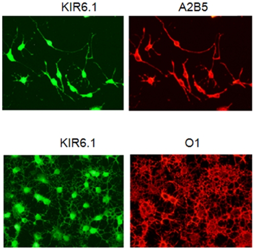
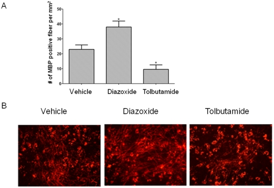
Methodology/findings: Studies were performed using rat oligodendrocyte precursor cell (OPC) cultures, cerebellar slice cultures, and an in vivo model of PWMI in which newborn mice were exposed to chronic sublethal hypoxia (10% O(2)). We found that K(ATP) channel components Kir 6.1 and 6.2 and SUR2 were expressed in oligodendrocytes. Additionally, diazoxide potently stimulated OPC proliferation, as did other K(ATP) activators. Diazoxide also stimulated myelination in cerebellar slice cultures. We also found that diazoxide prevented hypomyelination and ventriculomegaly following chronic sublethal hypoxia.
Conclusions: These results identify KATP channel components in OLs and show that diazoxide can stimulate OL proliferation in vitro. Importantly we find that diazoxide can promote myelination in vivo and prevent hypoxia-induced PWMI.
Conflict of interest statement
Figures


Similar articles
-
Diazoxide promotes oligodendrocyte differentiation in neonatal brain in normoxia and chronic sublethal hypoxia.Brain Res. 2014 Oct 24;1586:64-72. doi: 10.1016/j.brainres.2014.08.046. Epub 2014 Aug 23. Brain Res. 2014. PMID: 25157906 Free PMC article.
-
Characterization of an ATP-sensitive K(+) channel in rat carotid body glomus cells.Respir Physiol Neurobiol. 2011 Aug 15;177(3):247-55. doi: 10.1016/j.resp.2011.04.015. Epub 2011 Apr 22. Respir Physiol Neurobiol. 2011. PMID: 21536154 Free PMC article.
-
Ontogeny of sulfonylurea-binding regulatory subunits of K(ATP) channels in the pregnant rat myometrium.Reproduction. 2011 Jul;142(1):175-81. doi: 10.1530/REP-10-0492. Epub 2011 Apr 28. Reproduction. 2011. PMID: 21527399
-
KATP channel mutations in congenital hyperinsulinism.Semin Pediatr Surg. 2011 Feb;20(1):18-22. doi: 10.1053/j.sempedsurg.2010.10.012. Semin Pediatr Surg. 2011. PMID: 21185999 Review.
-
Novel Approaches to the Treatment of Preterm White Matter Injury through Targeting Remyelination.Clin Perinatol. 2025 Jun;52(2):289-306. doi: 10.1016/j.clp.2025.02.005. Epub 2025 Mar 26. Clin Perinatol. 2025. PMID: 40350212 Review.
Cited by
-
K(ATP) channel opener diazoxide prevents neurodegeneration: a new mechanism of action via antioxidative pathway activation.PLoS One. 2013 Sep 11;8(9):e75189. doi: 10.1371/journal.pone.0075189. eCollection 2013. PLoS One. 2013. PMID: 24040400 Free PMC article.
-
Adverse and protective influences of adenosine on the newborn and embryo: implications for preterm white matter injury and embryo protection.Pediatr Res. 2011 Apr;69(4):271-8. doi: 10.1203/PDR.0b013e31820efbcf. Pediatr Res. 2011. PMID: 21228731 Free PMC article. Review.
-
CHRONIC NEONATAL DIAZOXIDE THERAPY IS NOT ASSOCIATED WITH ADVERSE EFFECTS.Online J Biol Sci. 2014 Jan 1;14(1):49-56. doi: 10.3844/ojbsci.2014.49.56. Online J Biol Sci. 2014. PMID: 25587244 Free PMC article.
-
Diazoxide for Neonatal Hyperinsulinemic Hypoglycemia and Pulmonary Hypertension.Children (Basel). 2022 Dec 21;10(1):5. doi: 10.3390/children10010005. Children (Basel). 2022. PMID: 36670556 Free PMC article. Review.
-
Emergent Prophylactic, Reparative and Restorative Brain Interventions for Infants Born Preterm With Cerebral Palsy.Front Physiol. 2019 Jan 28;10:15. doi: 10.3389/fphys.2019.00015. eCollection 2019. Front Physiol. 2019. PMID: 30745876 Free PMC article. Review.
References
-
- Muglia LJ, Katz M. The Enigma of Spontaneous Preterm Birth. N Engl J Med. 2010;362:529–535. - PubMed
-
- Bodensteiner JB, Johnsen SD. Magnetic resonance imaging (MRI) findings in children surviving extremely premature delivery and extremely low birthweight with cerebral palsy. J Child Neurol. 2006;21:743–747. - PubMed
-
- Wilson-Costello D, Friedman H, Minich N, Fanaroff AA, Hack M. Improved survival rates with increased neurodevelopmental disability for extremely low birth weight infants in the 1990s. Pediatrics. 2005;115:997–1003. - PubMed
-
- Volpe JJ. Neurobiology of periventricular leukomalacia in the premature infant. Pediatr Res. 2001;50:553–562. - PubMed
-
- Back SA. Perinatal white matter injury: the changing spectrum of pathology and emerging insights into pathogenetic mechanisms. Ment Retard Dev Disabil Res Rev. 2006;12:129–140. - PubMed
Publication types
MeSH terms
Substances
Grants and funding
LinkOut - more resources
Full Text Sources
Other Literature Sources
Medical
