

Acute pulmonary alveolar proteinosis due to exposure to cotton dust
- PMID: 20532003
- PMCID: PMC2876706
- DOI: 10.4103/0970-2113.56355
Acute pulmonary alveolar proteinosis due to exposure to cotton dust
Abstract
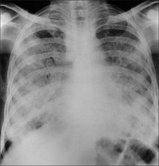
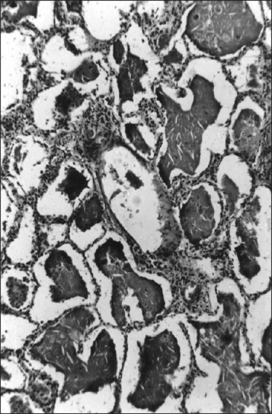
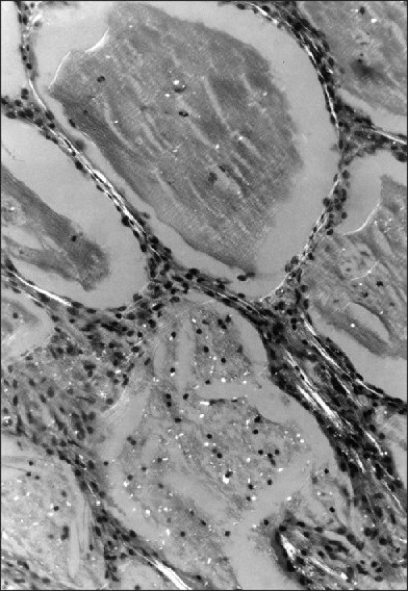
Secondary pulmonary alveolar proteinosis (PAP) is rare but may occur in association with malignancy, certain infections, and exposure to inorganic or organic dust and some toxic fumes. This case report describes the second recorded case of PAP due to exposure to cotton dust. A 24-year-old man developed PAP after working as a spinner for eight years without respiratory protection. He was admitted as an emergency patient with very severe dyspnea for four months and cough for several years. Chest X-ray showed bilateral diffuse alveolar consolidation. He died 16 days later, and a diagnosis of acute pulmonary alveolar proteinosis was made at autopsy. The histopathology demonstrated alveoli and respiratory bronchioles filled with characteristic periodic acid Schiff-positive material, which also revealed birefringent bodies of cotton dust under polarized light. Secondary PAP can be fatal and present with acute respiratory failure. The occupational history and characteristic pathology can alert clinicians to the diagnosis.
Keywords: Alveolar macrophage; bronchoalveolar lavage; cotton dust; granulocyte macrophage colony-stimulating factor; pulmonary alveolar proteinosis.
Conflict of interest statement
Figures
Similar articles
-
Combined-modality therapy for pulmonary alveolar proteinosis in a remote setting: a case report.BMC Pulm Med. 2019 Mar 12;19(1):61. doi: 10.1186/s12890-019-0822-x. BMC Pulm Med. 2019. PMID: 30866900 Free PMC article.
-
[Alveolar proteinosis after professional exposure to cotton and linen dust, successfully treated with whole lung lavage--a case report].Pneumonol Alergol Pol. 2004;72(5-6):217-20. Pneumonol Alergol Pol. 2004. PMID: 15757263 Polish.
-
Elevated Serum Anti-GM-CSF Antibodies before the Onset of Autoimmune Pulmonary Alveolar Proteinosis in a Patient with Sarcoidosis and Systemic Sclerosis.Tohoku J Exp Med. 2017 Sep;243(1):77-83. doi: 10.1620/tjem.243.77. Tohoku J Exp Med. 2017. PMID: 28966213
-
Pulmonary alveolar proteinosis: time to shift?Expert Rev Respir Med. 2015 Jun;9(3):337-49. doi: 10.1586/17476348.2015.1035259. Epub 2015 Apr 12. Expert Rev Respir Med. 2015. PMID: 25864717 Review.
-
Secondary pulmonary alveolar proteinosis in hematologic malignancies.Hematol Oncol Stem Cell Ther. 2014 Dec;7(4):127-35. doi: 10.1016/j.hemonc.2014.09.003. Epub 2014 Oct 6. Hematol Oncol Stem Cell Ther. 2014. PMID: 25300566 Review.
Cited by
-
"Crazy-paving" pattern: A characteristic presentation of pulmonary alveolar proteinosis and a review of the literature from India.Lung India. 2016 May-Jun;33(3):335-42. doi: 10.4103/0970-2113.180936. Lung India. 2016. PMID: 27186004 Free PMC article. No abstract available.
-
Clinical significance of cigarette smoking and dust exposure in pulmonary alveolar proteinosis: a Korean national survey.BMC Pulm Med. 2017 Nov 21;17(1):147. doi: 10.1186/s12890-017-0493-4. BMC Pulm Med. 2017. PMID: 29162083 Free PMC article.
-
Elemental analysis of lung tissue particles and intracellular iron content of alveolar macrophages in pulmonary alveolar proteinosis.Respir Res. 2011 Jun 30;12(1):88. doi: 10.1186/1465-9921-12-88. Respir Res. 2011. PMID: 21718529 Free PMC article.
-
Pulmonary alveolar proteinosis: Experience from a tertiary care center and systematic review of Indian literature.Lung India. 2016 Nov-Dec;33(6):626-634. doi: 10.4103/0970-2113.192876. Lung India. 2016. PMID: 27890991 Free PMC article.
-
Unsuspected pulmonary alveolar proteinosis in a patient with a slow resolving pneumonia: A case report.Respir Med Case Rep. 2013 Jun 26;10:1-3. doi: 10.1016/j.rmcr.2013.06.001. eCollection 2013. Respir Med Case Rep. 2013. PMID: 26029499 Free PMC article.
References
-
- Rosen RH, Castleman B, Liebow AA. Pulmonary alveolar proteinsos. N Engl J Med. 1958;258:1123–42. - PubMed
-
- Semour JF, Presneill JJ. Pulmonary alveolar proteinosis: State of the art. Am J Respir Crit Care Med. 2002;166:215–35. - PubMed
-
- Trapnell BC, Whitsett JA, Nakata K. Mechanism of disease: Pulmonary alveolar proteinosis. N Engl J Med. 2003;349:2527–39. - PubMed
-
- Ladeb S, Fleury-Faith J, Escadier Secondary alveolar proteinosis in cancer patients. Support Care Cancer. 1996;4:420–6. - PubMed
-
- Kosacka M, Dyla T, Jankowska R. Alveolar Proetinosis after Profession Exposure to cotton and linen dust, successful treated with whole lung lavage: A case report. Pneumonol Alergol Pol. 2004;72:217–20. - PubMed
LinkOut - more resources
Full Text Sources
Research Materials
