. 2010 Jun 4;5(6):e10967.
doi: 10.1371/journal.pone.0010967.

Prenatal inflammation-induced hypoferremia alters dopamine function in the adult offspring in rat: relevance for schizophrenia

Affiliations

Prenatal inflammation-induced hypoferremia alters dopamine function in the adult offspring in rat: relevance for schizophrenia

Argel Aguilar-Valles et al. PLoS One. .

Abstract

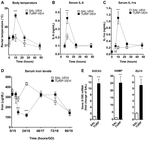
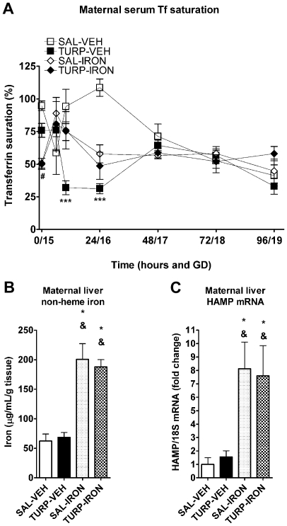
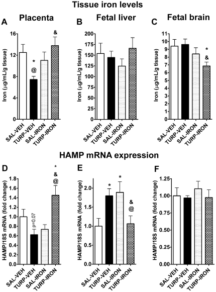
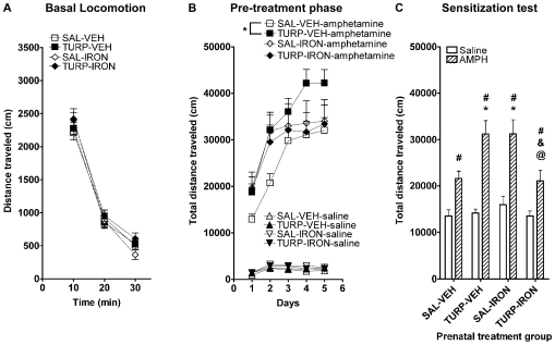
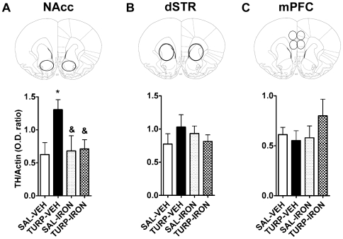
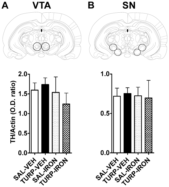
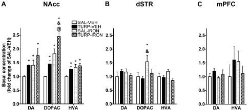
Maternal infection during pregnancy has been associated with increased incidence of schizophrenia in the adult offspring. Mechanistically, this has been partially attributed to neurodevelopmental disruption of the dopamine neurons, as a consequence of exacerbated maternal immunity. In the present study we sought to target hypoferremia, a cytokine-induced reduction of serum non-heme iron, which is common to all types of infections. Adequate iron supply to the fetus is fundamental for the development of the mesencephalic dopamine neurons and disruption of this following maternal infection can affect the offspring's dopamine function. Using a rat model of localized injury induced by turpentine, which triggers the innate immune response and inflammation, we investigated the effects of maternal iron supplementation on the offspring's dopamine function by assessing behavioral responses to acute and repeated administration of the dopamine indirect agonist, amphetamine. In addition we measured protein levels of tyrosine hydroxylase, and tissue levels of dopamine and its metabolites, in ventral tegmental area, susbtantia nigra, nucleus accumbens, dorsal striatum and medial prefrontal cortex. Offspring of turpentine-treated mothers exhibited greater responses to a single amphetamine injection and enhanced behavioral sensitization following repeated exposure to this drug, when compared to control offspring. These behavioral changes were accompanied by increased baseline levels of tyrosine hydroxylase, dopamine and its metabolites, selectively in the nucleus accumbens. Both, the behavioral and neurochemical changes were prevented by maternal iron supplementation. Localized prenatal inflammation induced a deregulation in iron homeostasis, which resulted in fundamental alterations in dopamine function and behavioral alterations in the adult offspring. These changes are characteristic of schizophrenia symptoms in humans.


Conflict of interest statement

Competing Interests: The authors have declared that no competing interests exist.

Figures

Figure 1
Figure 1. TURP induced a maternal inflammatory response and hypoferremia in pregnant rats at GD 15.
(A) Sterile saline (n = 6) or TURP (n = 8) was injected (i.m., 100 µl/rat) and core body temperature determined immediately before (0 time point) and 8, 10, 24 and 48 h after. TURP induced a significant increase in core body temperature 10 h after TURP injection, which receded 24 h after injection. p<0.0001: *** SAL vs. TURP (B and C) Serum IL-6 and IL-1ra levels were determined by ELISA from samples collected 6, 10, 24 and 48 h after SAL or TURP injections in the same rats as in (A). TURP induced a significant increased in the levels of both cytokines 10 h after injection and returned to baseline by 24 h. p<0.0001: *** SAL vs. TURP (D) Non-heme iron levels were also measured in the serum samples from the same animals used in (A-C). TURP induced significant reduction in non-heme levels 10 and 24 hours after TURP injection, returning to control levels by 48 hours. Non-heme iron levels decreased as pregnancy progressed, independently of treatment. p<0.0001: *** SAL vs. TURP (E) Hepatic gene expression in dams sacrificed at the peak of fever and cytokine induction, 10–11 hours after i.m. injection. SOCS3 (left panel), HAMP (middle panel) and zip14 (right panel) mRNAs were significantly induced in the TURP treated dams (n = 7 per group). p<0.0001: *** SAL vs. TURP.
Figure 2
Figure 2. TURP effect on maternal serum Tf saturation was prevented by parenteral iron supplementation.
(A) Serum Tf saturation levels were significantly reduced 10 and 24 h after TURP injection, this effect was absent in TURP-IRON treated dams. Baseline Tf saturation levels in iron supplemented animals was significantly reduced. (B and C) Hepatic non-heme iron levels, 96 h after i.m. injection, were unaffected by TURP treatment, but dramatically increased in iron supplemented dams. In parallel, iron supplementation led to a strong induction maternal hepatic HAMP mRNA expression. p<0.05 * vs. SAL-VEH, & vs. TURP-VEH.
Figure 3
Figure 3. Fetal non-heme iron and HAMP mRNA levels are modulated by maternal TURP treatment and iron supplementation.
(A) Non-heme iron levels in the placenta were lower in the TURP-treated dams, and this effect was reversed in the TURP-IRON treated dams. p<0.05: * vs. SAL-VEH, & vs. TURP-VEH, @ vs. SAL-IRON. SAL-VEH n = 6, TURP-VEH n = 8, SAL-IRON n = 7 and TURP-IRON n = 6. (B) Fetal liver non-heme iron content was unaffected by maternal treatments. (C) In the fetal brain, the only significant effect was a significant decrease in the non-heme iron content of TURP-IRON treated mothers. p<0.05: * vs. SAL-VEH, & vs. TURP-VEH. (D) Placental HAMP mRNA expression was decreased in TURP-VEH mothers, whereas this effect was significantly reversed in the TURP-IRON treated dams. p<0.05: * vs. SAL-VEH, & vs. TURP-VEH, @ vs. SAL-IRON. (E) In the fetal liver, HAMP mRNA expression was significantly increased by TURP and by iron supplementation alone, whereas when both treatments were given together these effects is blocked. p<0.05: * vs. SAL-VEH, & vs. TURP-VEH, @ vs. SAL-IRON. (F) HAMP mRNA expression was not significantly altered by any treatment in the fetal brain.
Figure 4
Figure 4. Prenatal TURP treatment increased acute and sensitized AMPH-induced locomotion, which are prevented by maternal iron co-administration.
(A) Basal locomotion in the adult offspring of SAL-VEH (n = 45), TURP-VEH (n = 50), SAL-IRON (n = 23) and TURP-IRON (n = 26) treated dams was not affected by any treatment when animals were introduced to the behavioral chambers for the first time. (B) AMPH administration (i.p. 2 mg/kg) for 5 consecutive days induced a progressive enhancement of the locomotor activating effects, which was overall grater among the offspring of TURP-VEH treated dams compare to their SAL-VEH counterparts. This effect was completely prevented by iron co-treated, with the offspring of the TURP-IRON and SAL-IRON groups showing identical levels of locomotor behavior, which were also indistinguishable from those of the SAL-VEH group. Saline pre-treated: SAL-VEH n = 24, TURP-VEH n = 26, SAL-IRON n = 12 and TURP-IRON n = 12. AMPH pre-treated: SAL-VEH n = 21, TURP-VEH n = 24, SAL-IRON n = 11 and TURP-IRON n = 14. (C) Seven days after the last saline or AMPH injection, all animals were tested for sensitization, with a lower dose of AMPH (i.p. 1 mg/kg). All AMPH pre-treated animals showed significantly greater response to this dose of AMPH (dashed bars) than the saline-pretreated animals (white bars). Among the AMPH pre-treated animals, those born to TURP-VEH and SAL-IRON presented significantly greater sensitization response, whereas in the offspring of TURP-IRON dams, sensitization response was comparable to control animals. p<0.01: # vs. saline pre-treated counterpart, * vs. AMPH pre-treated SAL-VEH, & vs. AMPH pre-treated TURP-VEH, @ vs. AMPH pre-treated SAL-IRON.
Figure 5
Figure 5. Iron supplementation prevented increased NAcc TH levels induced by prenatal TURP treatment.
(A) Basal TH levels in the NAcc were measured by western blotting from adult animals' protein extracts. These were significantly greater in the offspring of TURP-treated dams, which was prevented by maternal iron supplementation. SAL-VEH n = 5, TURP-VEH n = 7, SAL-IRON n = 5 and TURP-IRON n = 6. p<0.05 * vs. SAL-VEH, & vs. TURP-VEH. (B and C) TH protein levels from dSTR (B) and mPFC (C) were not significantly affected by any prenatal treatment.
Figure 6
Figure 6. TH expression in the somatodendritic DA regions is not affected by maternal treatments.
(A and B) TH protein levels were not significantly altered by maternal manipulations in VTA (A) and SN (B).
Figure 7
Figure 7. Prenatal inflammation and iron supplementation increase NAcc levels of DA, DOPAC and HVA.
(A) In the NAcc prenatal TURP induced a significant induction in the content of DA, DOPAC and HVA. These effects were also present in the offspring of SAL-IRON-treated dams and in the TURP-IRON offspring, who also showed DOPAC levels greater that all of the other groups. SAL-VEH n = 5, TURP-VEH n = 7, SAL-IRON n = 5 and TURP-IRON n = 6. p<0.05 * vs. SAL-VEH, & vs. TURP-VEH, @ vs. SAL-IRON. (B) In the dSTR only DOPAC content was significantly greater in the offspring of SAL-IRON dams, whereas no other significant effect was found on the metabolite or in DA and HVA levels. (C) In contrast, in the mPFC no changes were detected in DA and HVA (DOPAC was under detection limits of the assay for this region).

Similar articles

Cited by

References

    1. Brown AS, Derkits EJ. Prenatal Infection and Schizophrenia: A Review of Epidemiologic and Translational Studies. Am J Psychiatry. 2010. - PMC - PubMed
    1. Brown AS. Prenatal infection as a risk factor for schizophrenia. Schizophr Bull. 2006;32:200–202. - PMC - PubMed
    1. Babulas V, Factor-Litvak P, Goetz R, Schaefer CA, Brown AS. Prenatal exposure to maternal genital and reproductive infections and adult schizophrenia. Am J Psychiatry. 2006;163:927–929. - PubMed
    1. Sorensen HJ, Mortensen EL, Reinisch JM, Mednick SA. Association between prenatal exposure to bacterial infection and risk of schizophrenia. Schizophr Bull. 2009;35:631–637. - PMC - PubMed
    1. Cui K, Ashdown H, Luheshi GN, Boksa P. Effects of prenatal immune activation on hippocampal neurogenesis in the rat. Schizophr Res. 2009;113:288–297. - PubMed

Publication types