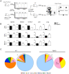
Naïve T cells re-distribute to the lungs of selectin ligand deficient mice
- PMID: 20532047
- PMCID: PMC2881108
- DOI: 10.1371/journal.pone.0010973
Naïve T cells re-distribute to the lungs of selectin ligand deficient mice
Abstract
Background: Selectin mediated tethering represents one of the earliest steps in T cell extravasation into lymph nodes via high endothelial venules and is dependent on the biosynthesis of sialyl Lewis X (sLe(x)) ligands by several glycosyltransferases, including two fucosyltransferases, fucosyltransferase-IV and -VII. Selectin mediated binding also plays a key role in T cell entry to inflamed organs.
Methodology/principal findings: To understand how loss of selectin ligands (sLe(x)) influences T cell migration to the lung, we examined fucosyltransferase-IV and -VII double knockout (FtDKO) mice. We discovered that FtDKO mice showed significant increases (approximately 5-fold) in numbers of naïve T cells in non-inflamed lung parenchyma with no evidence of induced bronchus-associated lymphoid tissue. In contrast, activated T cells were reduced in inflamed lungs of FtDKO mice following viral infection, consistent with the established role of selectin mediated T cell extravasation into inflamed lung. Adoptive transfer of T cells into FtDKO mice revealed impaired T cell entry to lymph nodes, but selective accumulation in non-lymphoid organs. Moreover, inhibition of T cell entry to the lymph nodes by blockade of L-selectin, or treatment of T cells with pertussis toxin to inhibit chemokine dependent G-coupled receptor signaling, also resulted in increased T cells in non-lymphoid organs. Conversely, inhibition of T cell egress from lymph nodes using FTY720 agonism of S1P1 impaired T cell migration into non-lymphoid organs.
Conclusions/significance: Taken together, our results suggest that impaired T cell entry into lymph nodes via high endothelial venules due to genetic deficiency of selectin ligands results in the selective re-distribution and accumulation of T cells in non-lymphoid organs, and correlates with their increased frequency in the blood. Re-distribution of T cells into organs could potentially play a role in the initiation of T cell mediated organ diseases.
Conflict of interest statement
Figures


Similar articles
-
Memory T cells are enriched in lymph nodes of selectin-ligand-deficient mice.J Immunol. 2010 Nov 15;185(10):5751-61. doi: 10.4049/jimmunol.1001878. Epub 2010 Oct 11. J Immunol. 2010. PMID: 20937846
-
Lack of functional selectin ligand interactions compromises long term tumor protection by CD8+ T cells.PLoS One. 2012;7(2):e32211. doi: 10.1371/journal.pone.0032211. Epub 2012 Feb 16. PLoS One. 2012. PMID: 22359671 Free PMC article.
-
Fucosyltransferase VII-deficient mice with defective E-, P-, and L-selectin ligands show impaired CD4+ and CD8+ T cell migration into the skin, but normal extravasation into visceral organs.J Immunol. 2002 Mar 1;168(5):2139-46. doi: 10.4049/jimmunol.168.5.2139. J Immunol. 2002. PMID: 11859099
-
FTY720 (fingolimod) in Multiple Sclerosis: therapeutic effects in the immune and the central nervous system.Br J Pharmacol. 2009 Nov;158(5):1173-82. doi: 10.1111/j.1476-5381.2009.00451.x. Epub 2009 Oct 8. Br J Pharmacol. 2009. PMID: 19814729 Free PMC article. Review.
-
Selectins and glycosyltransferases in leukocyte rolling in vivo.FEBS J. 2006 Oct;273(19):4377-89. doi: 10.1111/j.1742-4658.2006.05437.x. Epub 2006 Sep 5. FEBS J. 2006. PMID: 16956372 Review.
Cited by
-
Lack of functional selectin-ligand interactions enhances innate immune resistance to systemic Listeria monocytogenes infection.J Leukoc Biol. 2018 Feb;103(2):355-368. doi: 10.1002/JLB.4A1216-499R. Epub 2017 Dec 27. J Leukoc Biol. 2018. PMID: 29345354 Free PMC article.
-
Regulation of T-cell activation and migration by the kinase TBK1 during neuroinflammation.Nat Commun. 2015 Jan 21;6:6074. doi: 10.1038/ncomms7074. Nat Commun. 2015. PMID: 25606824 Free PMC article.
-
Lung-resident tissue macrophages generate Foxp3+ regulatory T cells and promote airway tolerance.J Exp Med. 2013 Apr 8;210(4):775-88. doi: 10.1084/jem.20121849. Epub 2013 Apr 1. J Exp Med. 2013. PMID: 23547101 Free PMC article.
-
Cutting edge: intravascular staining redefines lung CD8 T cell responses.J Immunol. 2012 Sep 15;189(6):2702-6. doi: 10.4049/jimmunol.1201682. Epub 2012 Aug 15. J Immunol. 2012. PMID: 22896631 Free PMC article.
-
Intravascular staining for discrimination of vascular and tissue leukocytes.Nat Protoc. 2014 Jan;9(1):209-22. doi: 10.1038/nprot.2014.005. Epub 2014 Jan 2. Nat Protoc. 2014. PMID: 24385150 Free PMC article.
References
-
- Lowe JB. Glycosylation, immunity, and autoimmunity. Cell. 2001;104:809–812. - PubMed
-
- Lowe JB. Glycan-dependent leukocyte adhesion and recruitment in inflammation. Curr Opin Cell Biol. 2003;15:531–538. - PubMed
-
- Lowe JB. Glycosylation in the control of selectin counter-receptor structure and function. Immunol Rev. 2002;186:19–36. - PubMed
-
- Yakubenia S, Wild MK. Leukocyte adhesion deficiency II. Advances and open questions. FEBS J. 2006;273:4390–4398. - PubMed
-
- Yakubenia S, Frommhold D, Scholch D, Hellbusch CC, Korner C, et al. Leukocyte trafficking in a mouse model for leukocyte adhesion deficiency II/congenital disorder of glycosylation IIc. Blood. 2008;112:1472–1481. - PubMed
Publication types
MeSH terms
Substances
Grants and funding
LinkOut - more resources
Full Text Sources
Molecular Biology Databases

