

Early postoperative pain and visual outcomes following epipolis-laser in situ keratomileusis and photorefractive keratectomy
- PMID: 20532139
- PMCID: PMC2882076
- DOI: 10.3341/kjo.2010.24.3.143
Early postoperative pain and visual outcomes following epipolis-laser in situ keratomileusis and photorefractive keratectomy
Abstract
Purpose: To compare early postoperative pain and visual outcomes after epipolis-laser in situ keratomileusis (epi-LASIK) and photorefractive keratectomy (PRK) in the treatment of myopia.
Methods: A retrospective chart review was designed and included 49 eyes in 30 patients who underwent epi-LASIK and 54 eyes in 29 patients who underwent PRK. During the early postoperative period (days 1 to 5), pain, uncorrected visual acuity (UCVA), and time to epithelial healing were recorded. Visual outcomes were followed for up to six months.
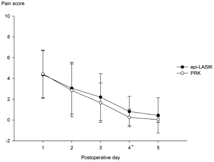
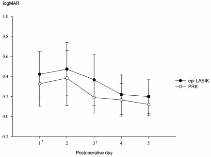
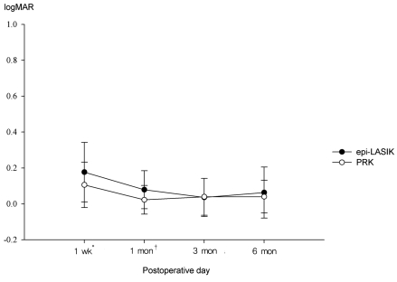
Results: Mean preoperative spherical equivalent refraction for the epi-LASIK group was -3.99+/-1.39 diopters (D) and that of the PRK group was -3.54+/-1.27 D. The pain scores on the fourth postoperative day were significantly higher in the epi-LASIK group than in the PRK group (p=0.017). Duration of pain in the epi-LASIK group was longer than in the PRK group (p=0.010). Mean healing time was significantly longer in the epi-LASIK group than in the PRK group (p<0.000). In addition, UCVA in the epi-LASIK group at postoperative days 1 and 3 were significantly lower than those in the PRK group (p=0.021 and p<0.000, respectively). Uncorrected visual acuity at one week and one month after epi-LASIK were lower than those after PRK (p=0.023 and p=0.004, respectively).
Conclusions: In the epi-LASIK patients, pain relief, corneal healing, and visual recovery seemed to be slower during the early postoperative period compared to those of the PRK patients. With longer duration of follow-up, however, there were no significant differences in visual outcome between the two groups.
Keywords: Epi-LASIK; Pain; Photorefractive keratectomy.
Conflict of interest statement
No potential conflict of interest relevant to this article was reported.
Figures
References
-
- Pallikaris IG, Naoumidi II, Kalyvianaki MI, Katsanevaki VJ. Epi-LASIK: comparative histological evaluation of mechanical and alcohol-assisted epithelial separation. J Cataract Refract Surg. 2003;29:1496–1501. - PubMed
-
- Pallikaris IG, Katsanevaki VJ, Kalyvianaki MI, Naoumidi II. Advances in subepithelial excimer refractive surgery techniques: Epi-LASIK. Curr Opin Ophthalmol. 2003;14:207–212. - PubMed
-
- Netto MV, Mohan RR, Ambrosio R, Jr, et al. Wound healing in the cornea: a review of refractive surgery complications and new prospects for therapy. Cornea. 2005;24:509–522. - PubMed
-
- Pallikaris IG, Kalyvianaki MI, Katsanevaki VJ, Ginis HS. Epi-LASIK: preliminary clinical results of an alternative surface ablation procedure. J Cataract Refract Surg. 2005;31:879–885. - PubMed
-
- Dai J, Chu R, Zhou X, et al. One-year outcomes of epi-LASIK for myopia. J Refract Surg. 2006;22:589–595. - PubMed
Publication types
MeSH terms
LinkOut - more resources
Full Text Sources
