

Suppressing glucose transporter gene expression in schistosomes impairs parasite feeding and decreases survival in the mammalian host
- PMID: 20532163
- PMCID: PMC2880588
- DOI: 10.1371/journal.ppat.1000932
Suppressing glucose transporter gene expression in schistosomes impairs parasite feeding and decreases survival in the mammalian host
Abstract
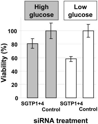
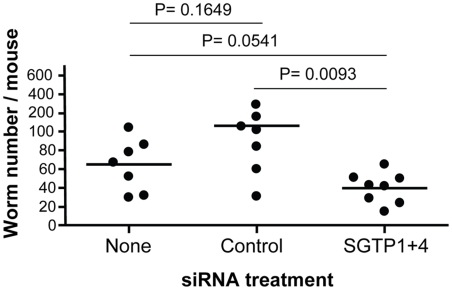
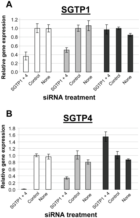
Adult schistosomes live in the host's bloodstream where they import nutrients such as glucose across their body surface (the tegument). The parasite tegument is an unusual structure since it is enclosed not by the typical one but by two closely apposed lipid bilayers. Within the tegument two glucose importing proteins have been identified; these are schistosome glucose transporter (SGTP) 1 and 4. SGTP4 is present in the host interactive, apical tegumental membranes, while SGTP1 is found in the tegumental basal membrane (as well as in internal tissues). The SGTPs act by facilitated diffusion. To examine the importance of these proteins for the parasites, RNAi was employed to knock down expression of both SGTP genes in the schistosomula and adult worm life stages. Both qRT-PCR and western blotting analysis confirmed successful gene suppression. It was found that SGTP1 or SGTP4-suppressed parasites exhibit an impaired ability to import glucose compared to control worms. In addition, parasites with both SGTP1 and SGTP4 simultaneously suppressed showed a further reduction in capacity to import glucose compared to parasites with a single suppressed SGTP gene. Despite this debility, all suppressed parasites exhibited no phenotypic distinction compared to controls when cultured in rich medium. Following prolonged incubation in glucose-depleted medium however, significantly fewer SGTP-suppressed parasites survived. Finally, SGTP-suppressed parasites showed decreased viability in vivo following infection of experimental animals. These findings provide direct evidence for the importance of SGTP1 and SGTP4 for schistosomes in importing exogenous glucose and show that these proteins are important for normal parasite development in the mammalian host.
Conflict of interest statement
The authors have declared that no competing interests exist.
Figures


Similar articles
-
Metabolite movement across the schistosome surface.J Helminthol. 2012 Jun;86(2):141-7. doi: 10.1017/S0022149X12000120. Epub 2012 Feb 27. J Helminthol. 2012. PMID: 22365312 Review.
-
Rapid appearance and asymmetric distribution of glucose transporter SGTP4 at the apical surface of intramammalian-stage Schistosoma mansoni.Proc Natl Acad Sci U S A. 1996 Apr 16;93(8):3642-6. doi: 10.1073/pnas.93.8.3642. Proc Natl Acad Sci U S A. 1996. PMID: 8622989 Free PMC article.
-
The role of tegumental aquaporin from the human parasitic worm, Schistosoma mansoni, in osmoregulation and drug uptake.FASEB J. 2009 Aug;23(8):2780-9. doi: 10.1096/fj.09-130757. Epub 2009 Apr 13. FASEB J. 2009. PMID: 19364765 Free PMC article.
-
[Immunocytochemical localization of two facilitated glucose transporters of Schistosoma mansoni in the tegument of Schistosoma japonicum].Zhongguo Ji Sheng Chong Xue Yu Ji Sheng Chong Bing Za Zhi. 1999;17(4):222-4. Zhongguo Ji Sheng Chong Xue Yu Ji Sheng Chong Bing Za Zhi. 1999. PMID: 12563769 Chinese.
-
Schistosoma mansoni: TGF-beta signaling pathways.Exp Parasitol. 2007 Nov;117(3):304-17. doi: 10.1016/j.exppara.2007.06.002. Epub 2007 Jun 16. Exp Parasitol. 2007. PMID: 17643432 Free PMC article. Review.
Cited by
-
Schistosome AMPK Is Required for Larval Viability and Regulates Glycogen Metabolism in Adult Parasites.Front Microbiol. 2021 Sep 1;12:726465. doi: 10.3389/fmicb.2021.726465. eCollection 2021. Front Microbiol. 2021. PMID: 34539616 Free PMC article.
-
RNA interference dynamics in juvenile Fasciola hepatica are altered during in vitro growth and development.Int J Parasitol Drugs Drug Resist. 2020 Dec;14:46-55. doi: 10.1016/j.ijpddr.2020.08.004. Epub 2020 Aug 19. Int J Parasitol Drugs Drug Resist. 2020. PMID: 32866764 Free PMC article.
-
Influence of Schistosoma japonicum programmed cell death protein 10 on the growth and development of schistosomula.Parasit Vectors. 2018 Jan 18;11(1):46. doi: 10.1186/s13071-018-2636-8. Parasit Vectors. 2018. PMID: 29347959 Free PMC article.
-
Schistosomes Impede ATP-Induced T Cell Apoptosis In Vitro: The Role of Ectoenzyme SmNPP5.Pathogens. 2022 Jan 26;11(2):155. doi: 10.3390/pathogens11020155. Pathogens. 2022. PMID: 35215099 Free PMC article.
-
A Review of Nanotechnology for Targeted Anti-schistosomal Therapy.Front Bioeng Biotechnol. 2020 Jan 31;8:32. doi: 10.3389/fbioe.2020.00032. eCollection 2020. Front Bioeng Biotechnol. 2020. PMID: 32083071 Free PMC article. Review.
References
-
- Morris GP, Threadgold LT. Ultrastructure of the tegument of adult Schistosoma mansoni. J Parasitol. 1968;54:15–27. - PubMed
-
- Fripp PJ. The sites of (1–14C) glucose assimilation in Schistosoma haematobium. Comp Biochem Physiol. 1967;23:893–898. - PubMed
-
- Rogers SH, Bueding E. Anatomical localization of glucose uptake by Schistosoma mansoni adults. Int J Parasitol. 1975;5:369–371. - PubMed
-
- Skelly P, Cunningham J, Kim J, Shoemaker C. Cloning, characterization and functional expression of cDNAs encoding glucose transporter proteins from the human parasite, Schistosoma mansoni. J Biol Chem. 1994;269:4247–4253. - PubMed
Publication types
MeSH terms
Substances
Associated data
- Actions
- Actions
Grants and funding
LinkOut - more resources
Full Text Sources
