. 2010 Jun 3;5(6):e10932.
doi: 10.1371/journal.pone.0010932.

Organization of the cpe locus in CPE-positive clostridium perfringens type C and D isolates

Affiliations

Organization of the cpe locus in CPE-positive clostridium perfringens type C and D isolates

Jihong Li et al. PLoS One. .

Abstract

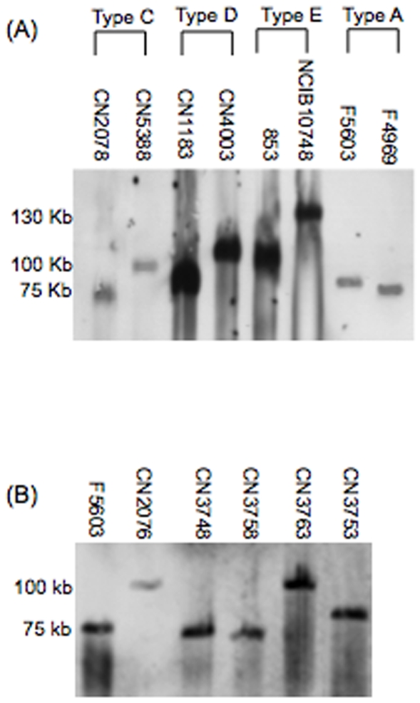
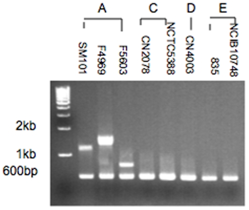
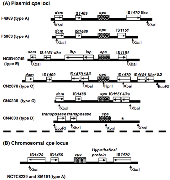
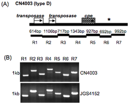
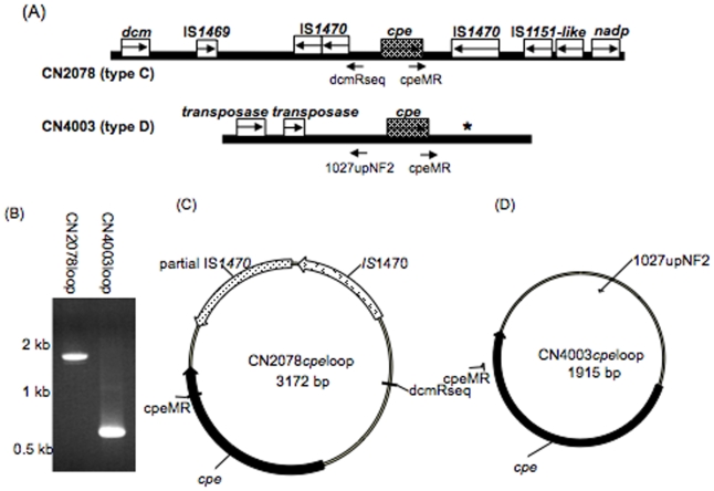
Clostridium perfringens enterotoxin (encoded by the cpe gene) contributes to several important human, and possibly veterinary, enteric diseases. The current study investigated whether cpe locus organization in type C or D isolates resembles one of the three (one chromosomal and two plasmid-borne) cpe loci commonly found amongst type A isolates. Multiplex PCR assays capable of detecting sequences in those type A cpe loci failed to amplify products from cpe-positive type C and D isolates, indicating these isolates possess different cpe locus arrangements. Therefore, restriction fragments containing the cpe gene were cloned and sequenced from two type C isolates and one type D isolate. The obtained cpe locus sequences were then used to construct an overlapping PCR assay to assess cpe locus diversity amongst other cpe-positive type C and D isolates. All seven surveyed cpe-positive type C isolates had a plasmid-borne cpe locus partially resembling the cpe locus of type A isolates carrying a chromosomal cpe gene. In contrast, all eight type D isolates shared the same plasmid-borne cpe locus, which differed substantially from the cpe locus present in other C. perfringens by containing two copies of an ORF with 67% identity to a transposase gene (COG4644) found in Tn1546, but not previously associated with the cpe gene. These results identify greater diversity amongst cpe locus organization than previously appreciated, providing new insights into cpe locus evolution. Finally, evidence for cpe gene mobilization was found for both type C and D isolates, which could explain their cpe plasmid diversity.


Conflict of interest statement

Competing Interests: The authors have declared that no competing interests exist.

Figures

Figure 1
Figure 1. PFGE cpe Southern blot analyses of cpe-positive type A, C, D and E isolates.
(A) DNA from type A, C, D or E isolates was subjected to PFGE prior to Southern blotting and hybridization with a DIG-labeled, cpe-specific probe. (B) DNA from type A (F5603) or type C (CN2076, CN3748, CN3758, CN3763 and CN3753) isolates was subjected to PFGE prior to Southern blotting and hybridization with a DIG-labeled, cpe-specific probe. The migration of molecular size markers is indicated on the left of the blot.
Figure 2
Figure 2. Analysis of cpe locus diversity in type C and D isolates using a multiplex PCR subtyping assay for cpe loci commonly found in type A isolates.
Representative results obtained with this assay are shown for type A isolates known to carry a chromosomal cpe gene (SM101), a plasmid cpe gene with an associated IS1470-like sequence (F4969), or a plasmid cpe gene with an associated IS1151 sequence (F5603). Also shown are representative results for this assay using culture lysates from cpe-positive type C isolates (CN2078, NCTC5388), cpe-positive type D isolates (CN4003) and type E isolates carrying silent cpe sequences (853 and NCIB10748). The migration of molecular size markers is indicated on the left of the blot.
Figure 3
Figure 3. RFLP analyses of cpe-positive type A, C, and D isolates and type E isolates carrying silent cpe sequences.
DNA from each isolate was digested with XbaI prior to conventional agarose gel electrophoresis and Southern blot hybridization with a cpe-specific probe. The migration of molecular weight markers is shown on the left of the blot.
Figure 4
Figure 4. Organization of cpe loci in type A, C, D and E.
A) Organization of plasmid cpe loci. B) Organization of the type A chromosome cpe locus. Each box represents an ORF. * indicates a region with sequence similarity to sequences present downstream of cpe in F4969, except for the absence of an IS1470-like gene. Sequences of cpe loci in F4969, F5603, NCIB10748, NCTC8239 and SM101 have been reported previously , , . Sequences of CN2078, CN5388 and CN4003 are based upon results of this study. The arrows show predicted enzyme (EcoRI, XbaI and KpnI) cleavage sites used in this study.
Figure 5
Figure 5. Overlapping PCR assay analysis of cpe locus diversity amongst type C isolates.
An overlapping PCR assay specific for amplification of the type C isolate CN2078 cpe locus region (R1 to R8) was performed using the primer battery shown in Table 2. (A) Map depicting the relationship between CN2078 cpe locus ORFs and reactions comprising this overlapping PCR battery. (B) PCR products produced by these reactions using DNA from type C isolates: CN2078, CN3753 and CN3748. (C) PCR products produced by these reactions using DNA from type C isolates: CN2076, CN3758 and CN3763. Numbers at left of each gel indicate migration of size markers in kb.
Figure 6
Figure 6. Overlapping PCR comparison of type C isolate CN5388 versus type A isolate F5603.
(A) Map depicting the relationship between ORFs and reactions in the cpe locus overlapping PCR battery (reactions R1 to R6) was performed using the primer battery show in Table 3. (B) Products of these reactions amplified by PCR using DNA from type C isolate CN5388 and CN2078 or type A isolates F5603. Arrows indicate that IS1151 sequences are oppositely oriented in CN5388 vs. F5603. (C) Products obtained when DNA from CN5388 or F5603 were subjected to a previously described overlapping PCR assay specific for the conserved region of type A cpe plasmids pCPF5603 and pCPF4969. Numbers at left of each gel indicate migration of size markers in kb.
Figure 7
Figure 7. Overlapping PCR assay analysis of cpe locus diversity amongst type D isolates.
An overlapping PCR assay specific for amplification of the type D isolate CN4003 cpe locus region (R1 to R7) was performed using the primer battery shown in Table 4. (A) Map depicting the relationship between ORFs and each reaction in this overlapping PCR battery. * indicates a region with sequence similarity to sequences downstream of cpe in F4969, except for the absence of IS1470-like gene. (B) Products of these reactions using DNA from two representative type D isolates: CN4003 and JGS1902. Numbers at left of each gel indicate migration of size markers in kb.
Figure 8
Figure 8. Detection of potential circular transposition intermediates carrying the cpe gene in type C and D isolates.
(A) Diagram of the cpe locus in type C isolate CN2078 and type D isolate CN4003. (B) PCR amplification of cpe-containing circular intermediates using the primers dcmRseq and cpeMR with CN2078 DNA or primers 1027upNF2 and cpeMR with CN4003 DNA. (C) Diagram derived from sequencing the CN2078 loop product of panel B that was amplified using primers dcmRseq and cpeMR. Black regions of the circle correspond to the amplified product. (D) Diagram derived from sequencing the product from CN4003 loop product of pane B that was amplified using primers 1027upNF2 and cpeMR. Black regions of the circle correspond to the amplified product.

Similar articles

Cited by

References

    1. McClane BA. Clostridium perfringens. In: Doyle MP, Beuchat LR, editors. Food Microbiology. 3rd ed. Washington D.C.: ASM press; 2007. pp. 423–444.
    1. McClane BA, Uzal FA, Miyakawa MF, Lyerly D, Wilkins T. The Enterotoxic Clostridia. In: Dworkin M, Falkow S, Rosenburg E, Schleifer H, Stackebrandt E, editors. The Prokaryotes. 3rd ed. New York: Springer NY; 2006. pp. 688–752.
    1. Kokai-Kun JF, Songer JG, Czeczulin JR, Chen F, McClane BA. Comparison of Western immunoblots and gene detection assays for identification of potentially enterotoxigenic isolates of Clostridium perfringens. J Clin Microbiol. 1994;32:2533–2539. - PMC - PubMed
    1. Bos J, Smithee L, McClane BA, Distefano RF, Uzal F, et al. Fatal necrotizing enteritis following a foodborne outbreak of enterotoxigenic Clostridium perfringens type A infection. Clin Infect Dis. 2005;15:78–83. - PubMed
    1. Collie RE, Kokai-Kun JF, McClane BA. Phenotypic characterization of enterotoxigenic Clostridium perfringens isolates from non-foodborne human gastrointestinal diseases. Anaerobe. 1998;4:69–79. - PubMed

Publication types

MeSH terms