. 2010 Jun 2;5(6):e10904.
doi: 10.1371/journal.pone.0010904.

Q344ter mutation causes mislocalization of rhodopsin molecules that are catalytically active: a mouse model of Q344ter-induced retinal degeneration

Affiliations

Q344ter mutation causes mislocalization of rhodopsin molecules that are catalytically active: a mouse model of Q344ter-induced retinal degeneration

Francis Concepcion et al. PLoS One. .

Abstract

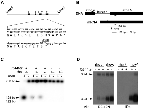
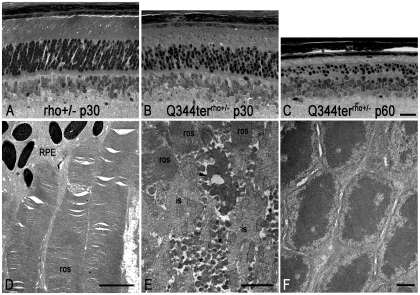
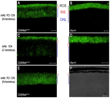
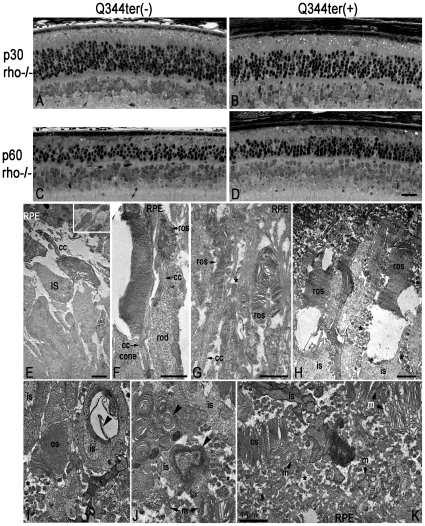
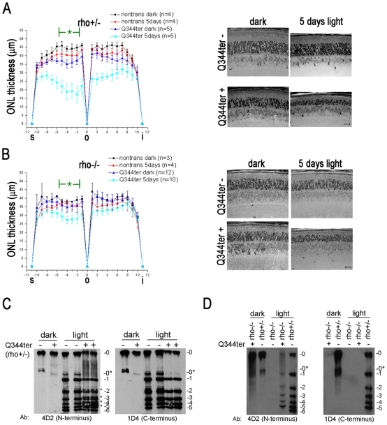
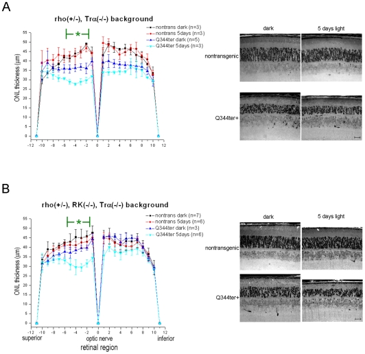
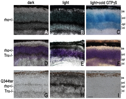
Q344ter is a naturally occurring rhodopsin mutation in humans that causes autosomal dominant retinal degeneration through mechanisms that are not fully understood, but are thought to involve an early termination that removed the trafficking signal, QVAPA, leading to its mislocalization in the rod photoreceptor cell. To better understand the disease mechanism(s), transgenic mice that express Q344ter were generated and crossed with rhodopsin knockout mice. Dark-reared Q344ter(rho+/-) mice exhibited retinal degeneration, demonstrating that rhodopsin mislocalization caused photoreceptor cell death. This degeneration is exacerbated by light-exposure and is correlated with the activation of transducin as well as other G-protein signaling pathways. We observed numerous sub-micrometer sized vesicles in the inter-photoreceptor space of Q344ter(rho+/-) and Q344ter(rho-/-) retinas, similar to that seen in another rhodopsin mutant, P347S. Whereas light microscopy failed to reveal outer segment structures in Q344ter(rho-/-) rods, shortened and disorganized rod outer segment structures were visible using electron microscopy. Thus, some Q344ter molecules trafficked to the outer segment and formed disc structures, albeit inefficiently, in the absence of full length wildtype rhodopsin. These findings helped to establish the in vivo role of the QVAPA domain as well as the pathways leading to Q344ter-induced retinal degeneration.


Conflict of interest statement

Competing Interests: The authors have declared that no competing interests exist.

Figures

Figure 1
Figure 1. Generation of Q344ter transgenic mice.
(A) Construct used to generate the Q344ter transgenic mice. In an 11-kb BamHI-flanked genomic clone containing the murine rod opsin gene, codon Q344 was mutated to an early stop signal (bottom *). The resulting rhodopsin mutant is missing the QVAPA domain but retains the six known potential phosphorylation sites (underlined). An AvrII site within the Q344ter transgene was generated by two silent mutations. Capitalized nucleotides denote the introduced point mutations. (B) Scheme to establish transcript expression level of the Q344ter transgene. The AvrII site is used to differentiate between Q344ter transgenic and WT transcript species. (C) Phosphor-image used to establish transgene-to-total rhodopsin transcript ratio. Total rhodopsin transcripts from each murine retina were amplified by RT-PCR and divided into two equal fractions. After AvrII digestion of one fraction, the enzyme-resistant 250 bp band is compared to the corresponding band from the undigested fraction. (D) Detection of the Q344ter mutant by western blot. Equal fraction (1/800) of a retina was loaded onto each lane. R2-12N monoclonal antibody recognizes the amino-terminus and identifies endogenous and Q344ter rhodopsin, while the 1D4 antibody recognizes only full length endogenous rhodopsin. Q344ter expression in rho−/− background was confirmed by R2-12N (left panel). As expected, these species were not detected by 1D4 (right panel). Monomeric and dimeric rhodopsin migrate at 33 kD and 66 kD, respectively.
Figure 2
Figure 2. Q344ter transgene causes retinal degeneration independent of light.
Images of retinal sections from epoxy-embedded eyecups were taken just above the optic nerve region from Q344terrho+/− (B, C) and their transgene-negative littermate control (A) mice at the indicated ages. All mice were born and reared in the dark. (D) Rod outer segment structure from control transgene-negative rho+/− mice. (E) Vesicular structures (asterisks) within the interphotoreceptor space of Q344terrho+/− retina. Arrow points to a degenerating structure. (F) Outer nuclear layer of Q344terrho+/− retina is devoid of vesicles. Scale bar in C (20 µm) is also representative for panels A and B. Scale bar  = 1 µm for D, E, and F. ros, rod outer segment; RPE, retinal pigmented epithelium.
Figure 3
Figure 3. Q344ter rhodopsin is mislocalized and does not support ROS formation when expressed in the absence of endogenous rhodopsin.
Q344terrho+/−, Q344rho−/− mice and their transgene negative littermate were dark-reared and sacrificed at p28–31. Frozen retinal sections immunostained with either the anti-N-terminal R2-12N (A, B, E), or the anti-C-terminal 1D4 (C, D) monoclonal antibodies against rhodopsin. R2-12N immunostaining was restricted to the rod outer segment (ROS) in control rho+/− sections (B), but extended to the rod inner segment (RIS) and outer nuclear layer (ONL) in Q344terrho+/− sections (A). 1D4 immunostaining revealed the presence of endogenous rhodopsin in the ONL in Q344terrho+/− retinas (C), which was not observed in retinas from negative transgene littermate controls (D). ROS structures were not detected in Q344terrho−/− retinal sections when stained with R2-12N (E) or imaged by DIC microscopy (F). Scale bars  = 20 µm.
Figure 4
Figure 4. Q344ter transgene does not accelerate retinal degeneration in rho−/− retinas.
As in Figure 2, these images of retinal sections from epoxy-embedded eyecups were taken just above the optic nerve region from Q344terrho−/− (B, D) and their transgene-negative littermate control (A, C) mice at p30 and p60. At both time points, ONL thicknesses appear similar in Q344terrho−/− mice and their littermate controls (compare A to B and C to D). Although progressive ONL thinning was observed in both groups, Q344ter does not appear to accelerate degeneration already occurring in rho−/− mice. Scale bar in D (20 µm) is representative for panels A–D. (E) Rho−/− rod photoreceptors do not elaborate outer segment structures. Instead, membrane tubules are seen (inset). The subretinal space is devoid of vesicular structures. Panels FH are from p30 Q344terrho−/− retinas, and IK are from p60 Q344terrho−/− retinas. (F) Short and disorganized rod outer segment can be seen distal to the connecting cilia. A neighboring cone photoreceptor with a much more intact outer segment is shown for comparison. (G) The rod outer segments shown here are thinner than normal (compare with Figure 2D). These structures are surrounded by apical processes from the RPE. (H) The membranous discs within some outer segment structures appear to be unstable. Numerous vesicular structures are present in the extracellular space (asterisks). (I-K) Vesicular structures are present in the interphotoreceptor space of Q344terrho−/− retinas. Outer segments containing discs are evident, but they are significantly compromised both in size and organization. Scale bars for E-K = 1 µm. Panels I-K are taken at the same magnification. os, outer segment; ros, rod outer segment; is, inner segment; cc, connecting cilium; RPE, retinal pigmented epithelium; m, membranous debris.
Figure 5
Figure 5. Light exacerbates Q344ter-induced retinal degeneration and activates Q344ter in the inner segment and outer nuclear layer.
(A) Q344terrho+/− and nontransgenic littermate control mice or (B) Q344terrho−/− and nontransgenic littermates control mice that were either dark-reared only or exposed to continuous light (3000 lux with undilated pupils) for five days were sacrificed at p28–31. Retinal sections near the optic nerve were analyzed by retinal morphometry. The diagram displays the mean (± SD) ONL thickness along the entire span of the retina. We focused on a light-sensitive region in the superior half near the optic nerve marked by a green asterisk where a slight ONL thinning occurred in light-exposed nontransgenic mice when compared to their dark-reared counterparts. Under dark-rearing, Q344ter retinas showed a moderate level of degeneration when compared to their transgene-negative littermate controls (p≤0.05). Light-exposure induced a severe form of degeneration in Q344ter transgenic retinas when compared to both light-exposed nontransgenic retinas and dark-reared Q344ter transgenic retinas (p≤0.05). A representative light microscopy image within this region from each group is displayed to the right. Scale bar  = 20 µm. (C) Isoelectric focusing gel of retinal extract from designated mice was blotted onto nitrocellulose and probed with the indicated antibodies against rhodopsin. The numbers to the right of each membrane image corresponds to the number of phosphates. In the left panel both Q344ter and WT rhodopsin molecules are detected by the anti-N-terminal rhodopsin mAb 4D2. Only non-phosphorylated rhodopsin (0) and apo-opsin (0*) species were detected in retinas from dark-reared mice. Light-exposure produced multiple phosphorylated rhodopsin species. Moreover, four extra bands (*) were detected in light-exposed Q344terrho+/− retinas. In the right panel, only WT rhodopsin molecules are detected by the anti-C-terminal rhodopsin mAb 1D4. Note that the four extra bands in the light-exposed Q344terrho+/− retinas detected by 4D2 are absent. (D) Mislocalized Q344ter molecules undergo light-dependent phosphorylation. With the rho−/− background, Q344ter molecules are mislocalized as shown by the absence of apparent ROS structures with R2-12N immunostaining (Fig. 3E). Retinal homogenates from light-exposed Q344terrho−/− mice were examined under similar IEF conditions described in (B). Light-dependent phosphorylation patterns of Q344ter molecules were detected by 4D2 (left panel) but not by 1D4 (right panel). This light-dependent phosphorylation pattern of Q344ter in these mice show that mislocalized Q344ter is capable of light-activation.
Figure 6
Figure 6. Light-exacerbated retinal degeneration in Q344ter mice is ameliorated in the Trα−/− background.
Dark-reared mice were kept in darkness or exposed to light for 5 days as in Fig. 5 (A) Retinal morphometry of Q344terrho+/− in the Trα−/− background. In the dark-reared group expression of the Q344ter transgene consistently caused a moderate thinning of the ONL. Light-exposure had no noticeable effect on the transgene negative mice, but caused further thinning of the ONL in Q344terrho+/− mice. However, this effect was less severe when compared to the Trα+/+ background (Fig. 5A). (B) The Q344terrho+/−, Trα−/− mice were further crossed into the RK−/− background to prevent light-induced formation of stable rhodopsin/Arr1 complexes. No additional rescuing effect beyond that seen in the Trα−/− background was observed in the light-exposed transgenic Q344ter mice in the RK−/−, Trα−/− background. Representative light micrographs of retinal sections are shown on the right. Scale bars  = 20 µm.
Figure 7
Figure 7. Light-dependent GTPγS loading (20 min exposure) in transgenic Q344ter frozen retinal sections.
[35S]GTPγS binding in situ was performed on unfixed frozen retinal sections from mice with the indicated genetic backgrounds. Basal [35S]GTPγS loading in the dark labels the synaptic layers (A, D, E), while light-exposure lead to additional labeling at the inner and outer segment compartments (B, E, H). Panels C, F, and I show non-specific background labeling. Scale bar  = 20 µm. All panels are taken at same magnification.

Similar articles

Cited by

References

    1. Rivolta C, Sharon D, DeAngelis MM, Dryja TP. Retinitis pigmentosa and allied diseases: numerous diseases, genes, and inheritance patterns. Hum Mol Genet. 2002;11:1219–1227. - PubMed
    1. Hamel C. Retinitis pigmentosa. Orphanet J Rare Dis. 2006;1:40. - PMC - PubMed
    1. Sohocki MM, Daiger SP, Bowne SJ, Rodriquez JA, Northrup H, et al. Prevalence of mutations causing retinitis pigmentosa and other inherited retinopathies. Hum Mutat. 2001;17:42–51. - PMC - PubMed
    1. Wang DY, Chan WM, Tam PO, Baum L, Lam DS, et al. Gene mutations in retinitis pigmentosa and their clinical implications. Clin Chim Acta. 2005;351:5–16. - PubMed
    1. Gregory-Evans K, Bhattacharya SS. Genetic blindness: current concepts in the pathogenesis of human outer retinal dystrophies. Trends Genet. 1998;14:103–108. - PubMed

Publication types